Small wounds leading to synovial infections

Operating on a synovial infection wound.jpg

Article by Peter Milner

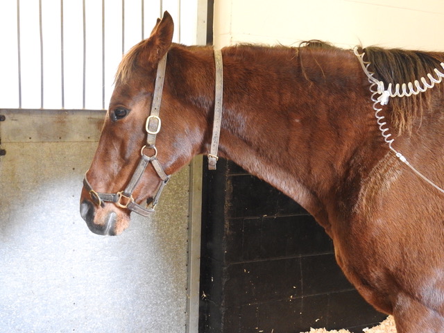
Most experienced trainers will know from bitter experience that a seemingly tiny wound can have a big impact if a horse is unlucky enough to sustain a penetrating injury right over a critical structure like a joint capsule or tendon sheath. Collectively, joints and tendon sheaths are called synovial structures, and synovial infection is a serious, potentially career-ending and sometimes life-threatening problem. 

A team of veterinary researchers from Liverpool University Veterinary School, published a study in Equine Veterinary Journal that examined factors influencing outcome and survival. This article was first published in European Trainer (issue 50 - summer 2015) but is being republished due to popular demand.

What is synovial infection?

Infection involving a synovial cavity, such as a joint or tendon sheath, is a common and potentially serious injury for the horse. The most prevalent cause is a wound, although a smaller proportion of cases result following an injection into a joint or tendon sheath, or after elective orthopaedic surgery to the area. Additionally, infection can occur via the bloodstream, particularly in foals that have not received enough colostrum.  Left untreated, the horse will remain in pain, and ongoing infection and inflammation can result in permanent damage. This can ultimately result in euthanasia on welfare grounds. 

What factors are important for horse survival?

synovial infections in Thoroughbred's.jpg

When a synovial infection occurs there is a huge inflammatory response, leading to swelling and pain. The horse usually shows severe lameness but following a good clinical examination, the cause is often quickly identified.  Prompt veterinary recognition of involvement of a joint or tendon sheath and aggressive treatment (involving flushing the affected synovial cavity and the correct use of systemic and local antibiotics) will often result in a good outcome for the horse.  Flushing removes inflammatory debris including destructive enzymes and free radicals, and it eliminates contaminating bacteria in most cases. This is performed most effectively by arthroscopic guidance (“keyhole” surgery) under general anaesthesia. Using a “scope” to do this is considered superior to flushing through needles because arthroscopy allows the inside of the problem area to be inspected, foreign material (for example, dirt or splinters of wood) to be removed, and any concurrent damage (such as damage to the cartilage or a cut into a tendon) to be evaluated. In addition, targeted high volume lavage is best achieved via arthroscopy. 

Survival following arthroscopic treatment of synovial sepsis is good – approximately 80-90% of adult horses undergoing a flush are discharged from hospital.  In foals, however, the figure is much lower, at around 55%, and this likely due to complicating factors such as concurrent sepsis involving multiple organs.  Our study, recently published in Equine Veterinary Journal, investigated what factors might be involved in determining survival to hospital discharge in 214 horses undergoing arthroscopic treatment for synovial sepsis. We used statistical modelling to evaluate the interactions with different factors at three key time points during the management of the condition at Liverpool Veterinary School, one of the leading UK referral veterinary hospitals. Information collected on admission to the hospital included when the horse was last seen to be normal, the cause of the infection, the degree of lameness present, and the level of white blood cells and protein in synovial fluid collected from the infected joint or tendon sheath. These lab tests are an important method which veterinarians use to determine how severe the infection is. Additional data collected included whether the surgery was performed out-of-normal working hours, if foreign material was present, the amount of inflammation present in the area, and whether any additional cartilage or tendon damage was found at surgery. Post-operative information gathered included what the levels of white blood cells and protein were in the synovial fluid after surgery and whether the horse needed further surgical treatment.

All horses in this study were greater than six months old and the majority had sustained a wound that communicated with a joint or tendon sheath.  Eighty-six per cent of the 214 horses admitted to the hospital survived to hospital discharge.  Of the 31 horses that did not survive, 27 were euthanised due to persistent infection or lameness.

An angry, protein-soup

A high level of protein in the synovial fluid of the affected joint or tendon sheath on admission and levels that remained high after surgery were strongly associated with a poor outcome and loss of the horse.  Protein concentrations are normally fairly low in a normal joint or tendon sheath, but protein leaks into the synovial cavity from surrounding blood vessels when inflamed. Protein is also produced by cells in the synovial cavity when they are activated in response to a severe insult such as infection. Protein clots trap bacteria in the joint, making it harder to remove infection. The protein soup also includes lots of inflammatory mediators such as enzymes and signalling molecules, and these cause further inflammation, tissue damage, and sensitise pain receptors in the synovial cavity magnifying the inflammatory response and increasing the pain experienced by the horse. Unchecked, this angry, inflamed environment can result in cartilage degeneration, bone damage, and adhesion (scar) formation. This fits well with another observation from this study linking the presence of moderate or severe synovial inflammation at surgery as a negative factor for survival. 

Small wounds can lead to big trouble

Interestingly, horses presenting with an obvious wound (as opposed to a small penetrating injury or no visible wound) were more likely to survive to hospital discharge. This may be due to the injury being noticed earlier and hence prompting earlier veterinary intervention. Alternatively, open wounds may allow drainage of inflammatory synovial fluid and lessen the detrimental effects of increased pressure within the joint as well as reducing ongoing exposure to inflammatory mediators. This finding highlights the fact that trainers should act promptly when faced with a wound – it is easy to underestimate just how much damage may be going on under the surface.

arthroscopic treatment of synovial sepsis.jpg

Horses undergoing surgical treatment of a joint or tendon sheath infection out-of-hours (for example in the middle of the night) were three times less likely to survive to hospital. Often, horses with a synovial infection arrive stressed and painful and not in an ideal state for having an anaesthetic. Early identification of an infection and appropriate management is important but stabilisation of the horse and preparation for surgery appear to outweigh any perceived benefits of undertaking immediate surgery.  This is borne out by the finding that time from initial injury to treatment was not associated with outcome and is in agreement with previous findings from other researchers. It is important to reiterate that prompt recognition and treatment of a horse with an infection in a synovial cavity is essential but that surgical management within 12-24 hours of diagnosis, so that the horse is in the best condition for undergoing anaesthesia, does not affect outcome. 

Do horses return to work after a synovial infection?

arthroscopic treatment of synovial sepsis.jpg

The big question that owners and trainers want to know is whether the horse will regain full function of the joint or tendon sheath after having an infection. Figures for return to function following surgical (arthroscopic) treatment for a synovial infection vary between 54-81%.  Various factors appear to relate to outcome but when looking at a predominately thoroughbred racing population, the statistic for return to training appears to be at the higher end of this range. Factors associated with failure to return to athletic performance include the presence of thickened inflammatory tissue (known as pannus) at the time of surgery and that may relate to the development of fibrous adhesions and scar tissue within joint or tendon sheath longer-term. Some structures are particularly likely to compromise future function, and horses with an infection of the navicular bursa in the foot following a nail penetration generally do worse. 

Take home message

Horses sustaining an infection to a joint or tendon sheath have a good chance of the infection clearing up and surviving the injury, with the likelihood of racing as high as around 80%.  Our key message for trainers from this study is that it is essential that they recognise early when an infection involves one of these structures and have a veterinarian fully evaluate the injury. Aggressive treatment is important and involves flushing the synovial cavity using a “scope” under anaesthesia to remove as much inflammatory and infective debris as possible. 

Equine Veterinary Journal - Wiley online library.jpg

Equine Pain: how can we recognise it and which painkiller should we use?

How well do we recognise pain in horses?We can all agree that alleviating pain in our patients is an important goal, but we may not be as good as we might hope at recognising pain in horses. Studies have shown that there is considerable variation in…
Screen Shot 2019-06-19 at 17.11.25.png

By Professor Celia Marr

We can all agree that alleviating pain in our patients is an important goal, but we may not be as good as we might hope at recognising pain in horses. Studies have shown that there is considerable variation in the scores vets assign when asked to predict how much pain they expect to see with specific clinical conditions.


Acute severe pain is perhaps most easily recognised by horsemen and vets; signs of severe colic, such as rolling, are usually very obvious. Low-grade pain, and pain not associated with abdominal disease can be more difficult to detect and go unrecognised. In particular, intra-thoracic pain and pain associated with injuries to the thoracic cage, withers and spine can be difficult to pinpoint.

This horse is clearly showing signs of abdominal pain—colic. It is lying down, has been rolling and is looking at its flank.

This horse is clearly showing signs of abdominal pain—colic. It is lying down, has been rolling and is looking at its flank.

Comfortable horses interact with their environment, look out over their stable door and eat willingly. Reluctance to move and restlessness indicate pain while looking at the flank, and kicking at the abdomen all suggest localised pain. Behaviours such as lifting hindlimbs, extending head, lateral and/or vertical head movements and pawing are also observed in uncomfortable horses.

Facial expression and pain

In humans, facial expressions are an important part of nonverbal communication. The Horse Grimace Scale has been developed to help identify subtle pain in horses. The grimace scale is easy to learn, can be applied quickly and takes into account our natural human tendency to focus on the face when evaluating both human and non-humans around us. This scale looks at ear position, tension around the eyes, tension in the chewing muscles and shape of the nostrils which tend to be held in a strained position if in pain. More complex pain scales incorporate facial expression with head position, flehmen, yawning, teeth grinding and interaction with people.

These scales were used in a recent Equine Veterinary Journal article looking at optimal methods to provide anaesthesia for castration. But, the focus on a strained facial expression, ears held back and lack of interaction with people can easily be misinterpreted as poor temperament. It is well worth trainers taking time to make sure their staff are educated on how to recognise signs of pain, as these sorts of clinical signs might indicate important conditions such as gastric ulcers, pneumonia or even musculoskeletal conditions such as fractured ribs. Yard staff should be encouraged to give horses the benefit of the doubt and report any apparent poor temperament so that veterinary investigations can be undertaken to get to the bottom of the problem. Similarly, these signs can be used to monitor horses after potentially painful procedures such as following surgery or castration.

What do we know about analgesic use in equine practice?

There is an increasingly large number of painkillers, also known as analgesics, which are either licensed for use in the horse or supported by research evidence. But it is likely that most equine vets use a relatively small range. British Equine Veterinary Association (BEVA) has recently tasked a team of its members to look at the evidence with underpin best practice for selections of analgesics in common clinical scenarios. This group is chaired by Professor Mark Bowen of the University of Nottingham and has been working for two years now and has collected evidence from the veterinary literature; and in parallel the group has consulted BEVA members to develop robust recommendations. The BEVA Clinical Practice Guidelines report on analgesia will be published soon and looks at the most effective analgesia in horses undergoing routine castration, horses with acute colic, orthopaedic pain and in horses with chronic pain that does not respond to standard non-steroidal anti-inflammatory drugs (NSAIDs) such as phenylbutazone (aka “Bute”). In making their recommendations around use of analgesics in horses, the BEVA team considered both the effectiveness of each analgesic drug, its safety and potential for side-effects.

What are the desirable characteristics of analgesic drugs?

The ideal analgesic has predictable effect and duration, minimal side effects and is easy to prescribe, purchase and administer, lacking any impact on the horse’s future status for human consumption. Of course, the ideal analgesic does not exist. To a large extent, the most appropriate analgesic will be dictated by the specific clinical indication.

Analgesia in colic

With colic, predictable level of analgesia and duration of action are key characteristics. The BEVA team found moderate evidence that flunixin provides superior analgesia to meloxicam and phenylbutazone in horses with colic. However, effective analgesia is desirable but very potent drugs are usually avoided for fear of masking declining clinical status in a horse which would be best served by surgical exploration rather than controlled with extremely potent analgesics. Potential damage to the gastrointestinal tract and effects on gastrointestinal motility are critical and the impact of concurrent shock and volume depletion must be considered. Similar considerations come into play with peri-operative pain but here, the level of analgesia required may be modified by the exact surgical indication and specific procedure and with some procedures, it will be appropriate to provide very potent analgesia, for example with surgical repair of fractures or other painful orthopaedic surgeries. In these cases, multimodal analgesia may well be indicated.

Analgesia following castration

The BEVA team found robust evidence to support a recommendation that pre-operative NSAIDs should be administered prior to surgery. They also recommended that analgesia should be given for at least three days after surgery and that local anaesthetic should be infused into the testicle even when a general anaesthetic is administered. Finally, they counselled that pre-operative use of butorphanol (a commonly used component of sedative protocols) alone should not be considered adequate analgesia for horses undergoing castration.

Selection of NSAIDs for musculoskeletal pain

Feed with phenylbutazone

Feed with phenylbutazone

There is moderate evidence to indicate that phenylbutazone provides superior analgesia for hoof pain / laminitis, compared to firocoxib and meloxicam but strong evidence to show that Suxibuzone can be used as a direct replacement for phenylbutazone in chronic orthopaedic pain. The evidence supporting the use of other NSAIDs is less definitive. Meloxicam and firocoxib may be equivalent to phenylbutazone for pain associated with inflammation of the joint lining and, although studies are not conclusive, the group came to the conclusion that ketoprofen is not as effective for addressing musculoskeletal pain.

One of the key safety recommendations relating to the use of phenylbutazone was that it is the NSAID that is most likely to induce gastrointestinal adverse events (right dorsal colitis or gastric glandular ulceration). However, although other NSAIDs have less adverse intestinal effects but can all be considered as potentially ulcerogenic. Horses on long-term analgesic therapy should be monitored carefully and further investigations undertaken if they show weight loss, poor appetite or develop the more general signs of pain described above, as this might indicate that the NSAID is having adverse effects on the intestine.

The BEVA team also concluded that giving NSAIDs at doses above those generally recommended in veterinary texts and stacking (i.e., combining maximal doses of different NSAIDs) should be avoided. These practices simply increase risk with no analgesic benefit.

Alternative analgesics

Unfortunately, there are not many practical alternatives to NSAIDs. There is research ongoing looking at topical NSAIDs and alternative drugs such as paracetamol, tramadol and fentanyl. These drugs are unlikely to be in common use in horses in training but do have a place in management of horses with more severe clinical problems.

Similarly, morphine and methadone are used commonly in equine hospitals, but these Schedule 2 controlled drugs are generally not used widely in practice. Buprenorphine has been extensively researched recently and evidence is accumulating supporting its use particularly in the peri-operative patient.

Final warnings

The BEVA group’s report contained a clear warning that highly potent analgesia should only be utilised under the direct control of a veterinary surgeon who has fully evaluated a horse and having developed a therapeutic, analgesic plan that includes ongoing monitoring. It is also important to bear in mind that the best way to alleviate pain associated with a specific clinical condition is to cure the underlying cause. Painkillers should always be used with respect and not be seen as a way to patch up a horse that has an undiagnosed musculoskeletal problem or internal condition.

BUY THIS ISSUE IN PRINT OR DOWNLOAD

July - September 2019, issue 66 (PRINT)
£8.95
Quantity:
Add to Cart

WHY NOT SUBSCRIBE?

DON'T MISS OUT AND SUBSCRIBE TO RECEIVE THE NEXT FOUR ISSUES!

Equine Herpesvirus-1 : An Elusive Target

Equine herpesvirus -1; an elusive targetInfectious diseases are not uncommon in racehorses in training, breeding stock, and pleasure horses. Some of the more serious diseases can be financially devastating to the animal’s owners and to the equine in…

By Neil Bryant 

Infectious diseases are not uncommon in racehorses in training, breeding stock, and pleasure horses. Some of the more serious diseases can be financially devastating to the animal’s owners and to the equine industry on the whole. Viruses belonging to the herpesvirus family cause some of the most well characterized equine infectious diseases, and the most problematic of these is equine herpesvirus 1 (EHV-1; species Equid alphaherpesvirus 1). EHV-1 is ubiquitous in most horse populations in the world. It is responsible for major economic and welfare problems causing respiratory disease, neurological disease (mainly seen in adult horses), and abortion and neonatal foal death in pregnant mares.

This was most notably highlighted by the multiple abortion outbreak recorded in Hertfordshire, England, between February and April 2016 in fully vaccinated animals (http://www.aht.org.uk/cms-display/interim-report16-april2.html). Studies have determined that EHV-1 is a common cause of abortion. Occasional cases have also been linked to EHV-4 infection, but this is much rarer and doesn’t account for episodes of multiple abortion, as is seen occasionally with EHV-1.

The virus

EHV-1 was first isolated from an equine abortion in the U.S. in the 1930s. At the time of first isolation the vets weren’t sure what it was, but they knew it was infectious. Subsequent genetic analysis much later led to the classification of the virus in the genus Varicellovirus (family Herpesviridae), together with its close relatives equine herpesvirus 4 (EHV-4; species Equid alphaherpesvirus 4) and equine herpesvirus 8 (EHV-8; species Equid alphaherpesvirus 8). Interestingly it is grouped with, and is therefore genetically similar to, the human herpesvirus responsible for chickenpox, the Varicella Zoster virus. Initial infection of horses was thought to occur around weaning, when virus-neutralizing antibodies transferred to the foal from the mare’s colostrum had declined enough to make them susceptible to infection. However, virus has been isolated from foals as young as seven days old with high antibody levels but without any significant clinical signs. Immunity to re-infection after primary infection is relatively short-lived, lasting between three-six months, but it is rare for naturally infected mares to abort in consecutive pregnancies.

TO READ MORE --

BUY THIS ISSUE IN PRINT OR DOWNLOAD -

April - June 2018, issue 61 (PRINT)
£8.95
Quantity:
Add to Cart

Why not subscribe? 

Don't miss out and subscribe to receive the next four issues!