Small wounds leading to synovial infections

Article by Peter Milner
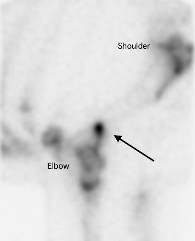
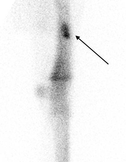
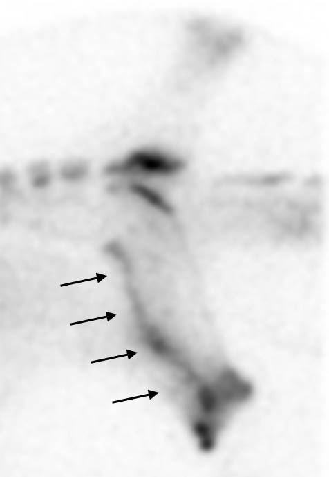
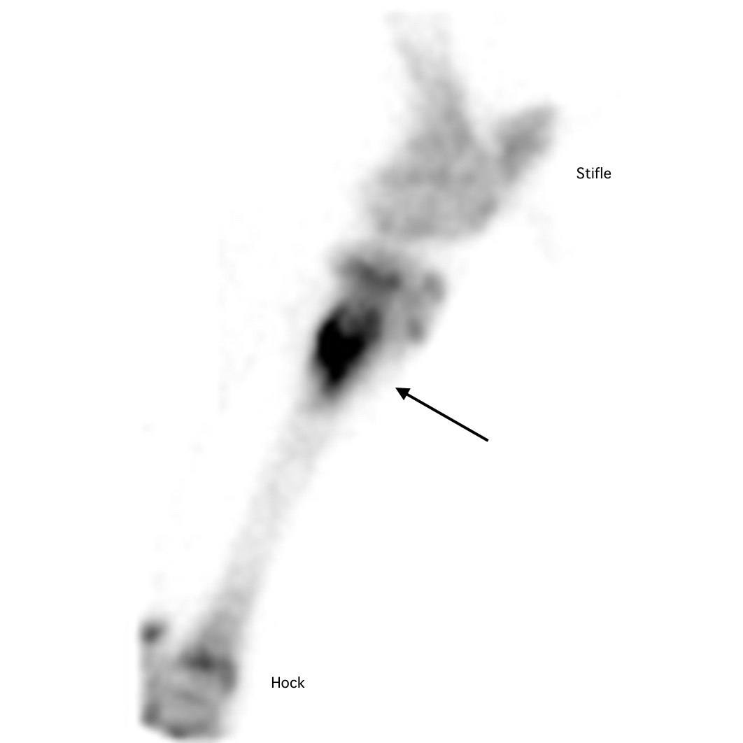
Most experienced trainers will know from bitter experience that a seemingly tiny wound can have a big impact if a horse is unlucky enough to sustain a penetrating injury right over a critical structure like a joint capsule or tendon sheath. Collectively, joints and tendon sheaths are called synovial structures, and synovial infection is a serious, potentially career-ending and sometimes life-threatening problem.
A team of veterinary researchers from Liverpool University Veterinary School, published a study in Equine Veterinary Journal that examined factors influencing outcome and survival. This article was first published in European Trainer (issue 50 - summer 2015) but is being republished due to popular demand.
What is synovial infection?
Infection involving a synovial cavity, such as a joint or tendon sheath, is a common and potentially serious injury for the horse. The most prevalent cause is a wound, although a smaller proportion of cases result following an injection into a joint or tendon sheath, or after elective orthopaedic surgery to the area. Additionally, infection can occur via the bloodstream, particularly in foals that have not received enough colostrum. Left untreated, the horse will remain in pain, and ongoing infection and inflammation can result in permanent damage. This can ultimately result in euthanasia on welfare grounds.
What factors are important for horse survival?

When a synovial infection occurs there is a huge inflammatory response, leading to swelling and pain. The horse usually shows severe lameness but following a good clinical examination, the cause is often quickly identified. Prompt veterinary recognition of involvement of a joint or tendon sheath and aggressive treatment (involving flushing the affected synovial cavity and the correct use of systemic and local antibiotics) will often result in a good outcome for the horse. Flushing removes inflammatory debris including destructive enzymes and free radicals, and it eliminates contaminating bacteria in most cases. This is performed most effectively by arthroscopic guidance (“keyhole” surgery) under general anaesthesia. Using a “scope” to do this is considered superior to flushing through needles because arthroscopy allows the inside of the problem area to be inspected, foreign material (for example, dirt or splinters of wood) to be removed, and any concurrent damage (such as damage to the cartilage or a cut into a tendon) to be evaluated. In addition, targeted high volume lavage is best achieved via arthroscopy.
Survival following arthroscopic treatment of synovial sepsis is good – approximately 80-90% of adult horses undergoing a flush are discharged from hospital. In foals, however, the figure is much lower, at around 55%, and this likely due to complicating factors such as concurrent sepsis involving multiple organs. Our study, recently published in Equine Veterinary Journal, investigated what factors might be involved in determining survival to hospital discharge in 214 horses undergoing arthroscopic treatment for synovial sepsis. We used statistical modelling to evaluate the interactions with different factors at three key time points during the management of the condition at Liverpool Veterinary School, one of the leading UK referral veterinary hospitals. Information collected on admission to the hospital included when the horse was last seen to be normal, the cause of the infection, the degree of lameness present, and the level of white blood cells and protein in synovial fluid collected from the infected joint or tendon sheath. These lab tests are an important method which veterinarians use to determine how severe the infection is. Additional data collected included whether the surgery was performed out-of-normal working hours, if foreign material was present, the amount of inflammation present in the area, and whether any additional cartilage or tendon damage was found at surgery. Post-operative information gathered included what the levels of white blood cells and protein were in the synovial fluid after surgery and whether the horse needed further surgical treatment.
All horses in this study were greater than six months old and the majority had sustained a wound that communicated with a joint or tendon sheath. Eighty-six per cent of the 214 horses admitted to the hospital survived to hospital discharge. Of the 31 horses that did not survive, 27 were euthanised due to persistent infection or lameness.
An angry, protein-soup
A high level of protein in the synovial fluid of the affected joint or tendon sheath on admission and levels that remained high after surgery were strongly associated with a poor outcome and loss of the horse. Protein concentrations are normally fairly low in a normal joint or tendon sheath, but protein leaks into the synovial cavity from surrounding blood vessels when inflamed. Protein is also produced by cells in the synovial cavity when they are activated in response to a severe insult such as infection. Protein clots trap bacteria in the joint, making it harder to remove infection. The protein soup also includes lots of inflammatory mediators such as enzymes and signalling molecules, and these cause further inflammation, tissue damage, and sensitise pain receptors in the synovial cavity magnifying the inflammatory response and increasing the pain experienced by the horse. Unchecked, this angry, inflamed environment can result in cartilage degeneration, bone damage, and adhesion (scar) formation. This fits well with another observation from this study linking the presence of moderate or severe synovial inflammation at surgery as a negative factor for survival.
Small wounds can lead to big trouble
Interestingly, horses presenting with an obvious wound (as opposed to a small penetrating injury or no visible wound) were more likely to survive to hospital discharge. This may be due to the injury being noticed earlier and hence prompting earlier veterinary intervention. Alternatively, open wounds may allow drainage of inflammatory synovial fluid and lessen the detrimental effects of increased pressure within the joint as well as reducing ongoing exposure to inflammatory mediators. This finding highlights the fact that trainers should act promptly when faced with a wound – it is easy to underestimate just how much damage may be going on under the surface.

Horses undergoing surgical treatment of a joint or tendon sheath infection out-of-hours (for example in the middle of the night) were three times less likely to survive to hospital. Often, horses with a synovial infection arrive stressed and painful and not in an ideal state for having an anaesthetic. Early identification of an infection and appropriate management is important but stabilisation of the horse and preparation for surgery appear to outweigh any perceived benefits of undertaking immediate surgery. This is borne out by the finding that time from initial injury to treatment was not associated with outcome and is in agreement with previous findings from other researchers. It is important to reiterate that prompt recognition and treatment of a horse with an infection in a synovial cavity is essential but that surgical management within 12-24 hours of diagnosis, so that the horse is in the best condition for undergoing anaesthesia, does not affect outcome.
Do horses return to work after a synovial infection?

The big question that owners and trainers want to know is whether the horse will regain full function of the joint or tendon sheath after having an infection. Figures for return to function following surgical (arthroscopic) treatment for a synovial infection vary between 54-81%. Various factors appear to relate to outcome but when looking at a predominately thoroughbred racing population, the statistic for return to training appears to be at the higher end of this range. Factors associated with failure to return to athletic performance include the presence of thickened inflammatory tissue (known as pannus) at the time of surgery and that may relate to the development of fibrous adhesions and scar tissue within joint or tendon sheath longer-term. Some structures are particularly likely to compromise future function, and horses with an infection of the navicular bursa in the foot following a nail penetration generally do worse.
Take home message
Horses sustaining an infection to a joint or tendon sheath have a good chance of the infection clearing up and surviving the injury, with the likelihood of racing as high as around 80%. Our key message for trainers from this study is that it is essential that they recognise early when an infection involves one of these structures and have a veterinarian fully evaluate the injury. Aggressive treatment is important and involves flushing the synovial cavity using a “scope” under anaesthesia to remove as much inflammatory and infective debris as possible.

The On-going Effort to Minimise the Rate and Impact of Fractures
Published in European Trainer, January - March 2018, issue 60.
In thoroughbred racing, musculoskeletal injury is a major safety concern and is the leading reason for days lost to training. Musculoskeletal injury is the greatest reason for horse turnover in racing stables, with financial implications for the owner and the racing industry. Injuries, particularly on race day, have an impact on public perception of racing.
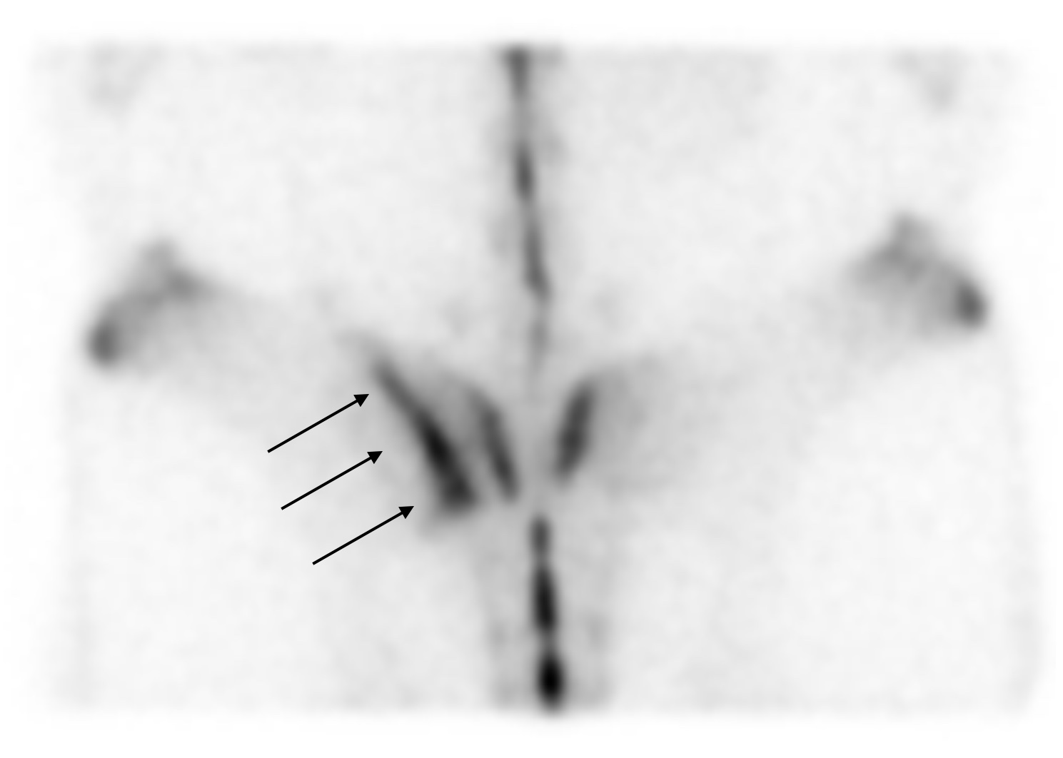
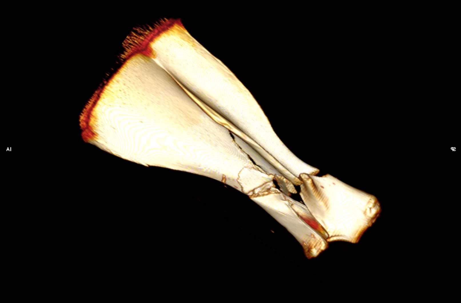
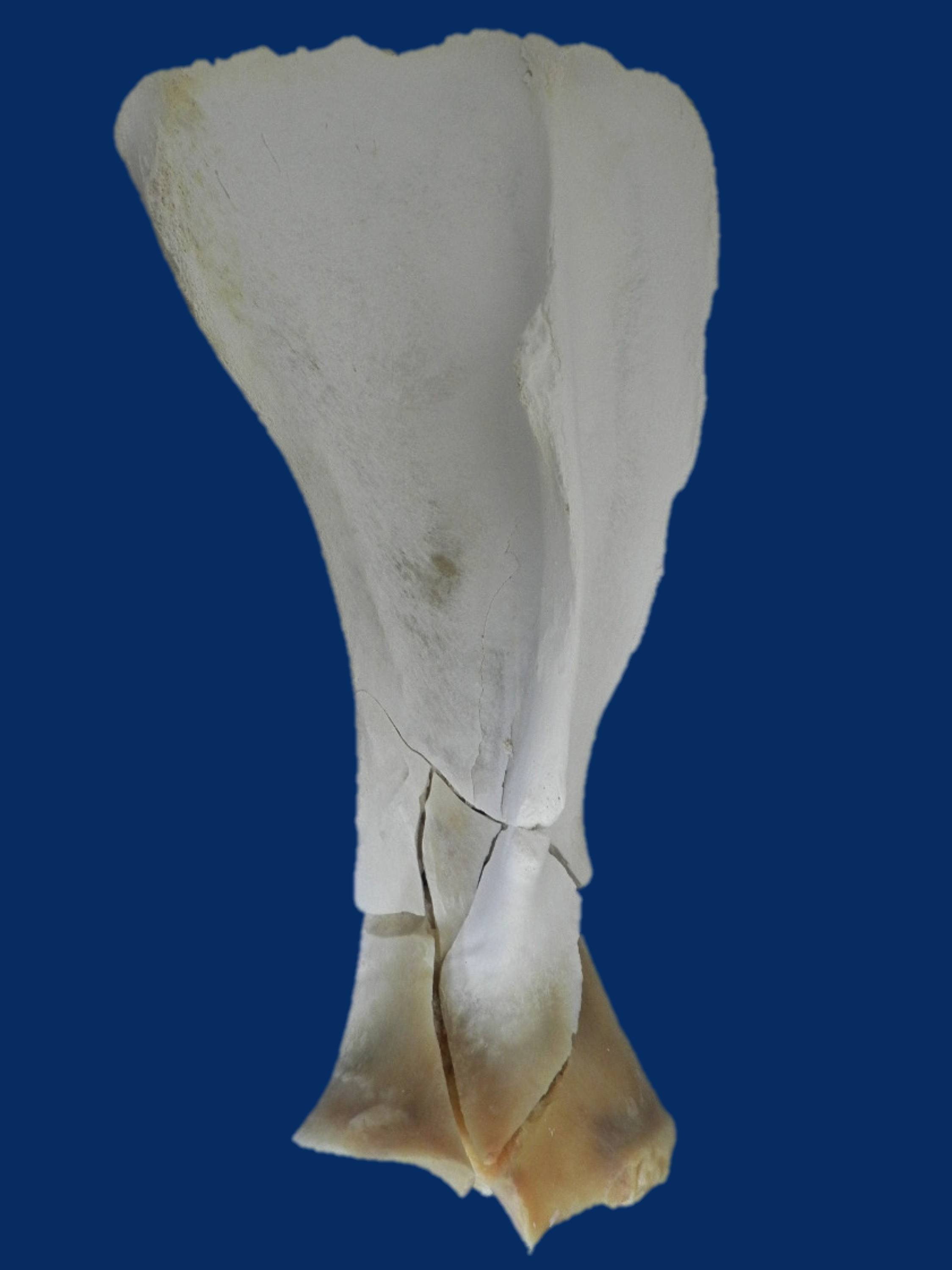
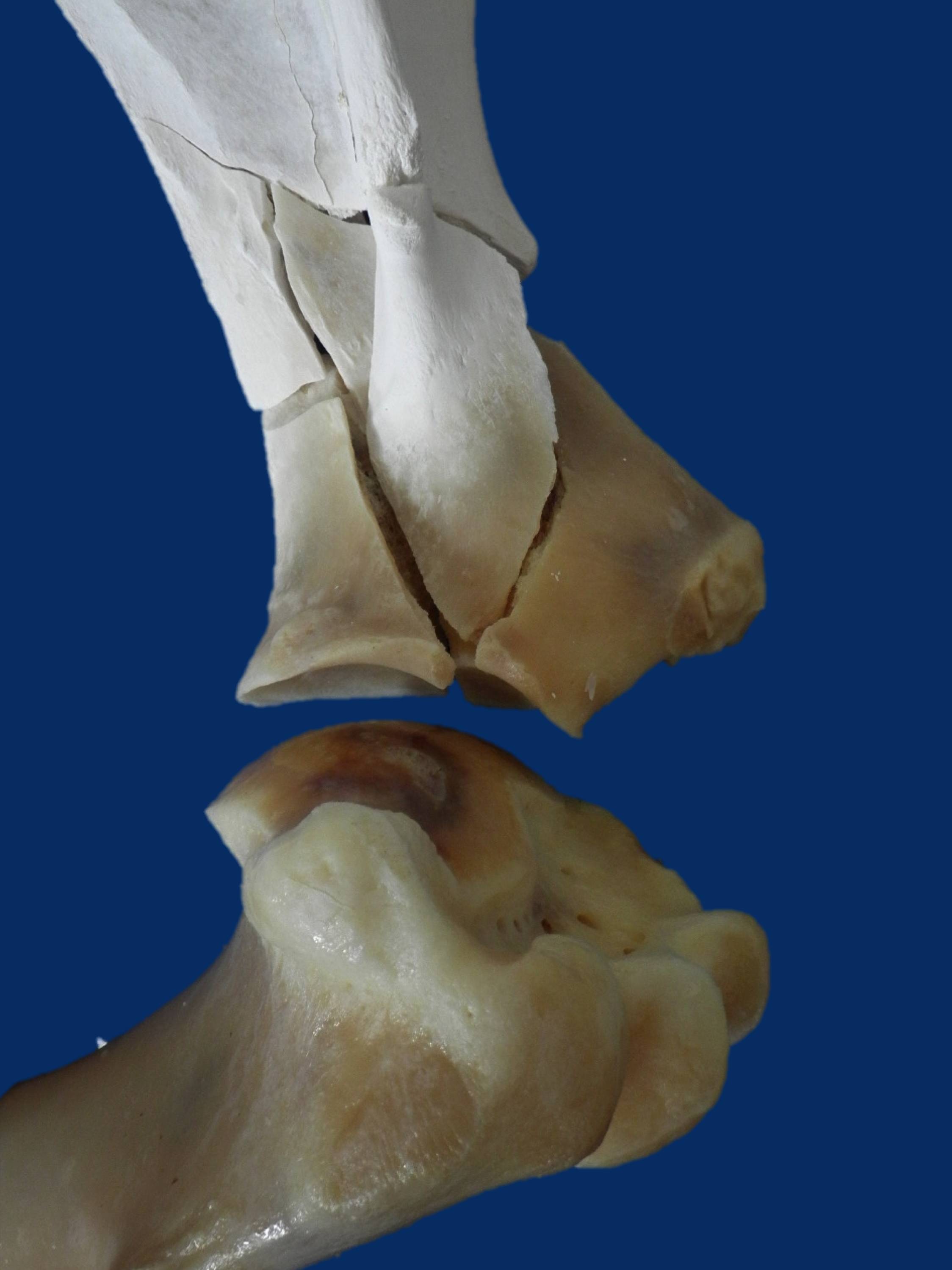
Upper limb and pelvis fractures are less common than lower limb fractures, but they can lead to fatalities. Reducing the overall prevalence of fractures is critical and, at the very least, improving the rate of detection of fractures in their early stages so the horse can be withdrawn from racing with a recoverable injury will be a big step forwards in racehorse welfare. Currently, we lack information on the outcomes following fracture, and an article recently published in the Equine Veterinary Journal (EVJ) from the veterinary team at the Hong Kong Jockey Club (HKJC) addressed this important knowledge gap.
Hong Kong Fracture Outcome Study
The HKJC veterinary team is in a unique position to carry out this work because their centralised and computerised database of clinical records, together with racing and retirement records, allows them to document follow-up, which is all but impossible elsewhere in the world. Dr Leah McGlinchey, working with vets in Hong Kong and researchers from the Royal Veterinary College, London, reviewed clinical records from 2003 to 2014 to identify racehorses that suffered a fracture or fractures to the bones of the upper limb or the pelvis during training or racing, confirmed by nuclear scintigraphy, radiography, ultrasonography, or autopsy....
To read more - subscribe now!
Gallery
A report from the Merial - Performance Horse CPD and Raceday at Gowran Park

Becky James BSc, MSc - Haygain
Published in European Trainer - October - December 2017, issue 59
Click here to order this back issue!
Vets from all over Ireland congregated at Gowran Park racecourse in July for a continuing professional development event on the Performance Horse. The event, organised by European Trainer Magazine and Merial Animal Health, was the second in a series of veterinary CPD events for 2017 and featured a panel of expert speakers. The event was co-sponsored by Haygain and Connolly’s RED MILLS.
Managing Inflammatory Airway Disease – Dr Emmanuelle Van Erck-Westergren
The first speaker Dr Van Erck-Westergren was due to fly in from Brussels on the morning of the event, so when her flight was cancelled at the last minute there was a moment of concern for the organisers but they arranged to bring her into the room via a video link so all was not lost!
Using her experience in practice at the Equine Sports Medicine Practice in Belgium, Dr Van Erck explained the importance of vets helping clients to manage the environment of the horses to prevent and manage Inflammatory Airway Disease (IAD). She described managing the horse’s environment to reduce exposure to noxious inhalable particles and improve hygiene and ventilation in the stable as the cornerstone to the success of treating IAD.
Important considerations for the environment include building design, bedding, stable activities and most critically, the forage, as this is in the horse’s breathing zone. Dr Van Erck explained that hay remains an important source of forage for horses but it is also a major source of dust and contaminants. Soaking hay is a cheap way of reducing airborne dust but it promotes bacterial proliferation and leaches out the nutritional value so well-made haylage or preferably steamed hay should only be fed to horses with IAD.
To read more of this report - subscribe here!
Hoof - Surface Interaction Study

By Amy Barstow BVetMed, MRCVS, PhD Candidate
First published in European Trainer issue 58 - July - September 2017
Click here to order this back issue!
In racehorses, the identification of risk factors for lameness has driven research efforts into lameness prevention strategies.
The foot-surface interaction is of particular interest and has been the subject of a lot of research. To date this has primarily been through the investigation of ground-surface composition and properties, in addition to the effects specific surfaces have on the horse’s way of going. Changes in speed, stride length, and frequency have been observed, and the vibrations (shock waves) resulting from the initial foot-surface collision have been measured on different surfaces. However, the surface represents only half the story: what about the foot? In practice it is the foot that we are more likely to influence on an individual level.
As part of my PhD studies at the Royal Veterinary College (RVC), we have investigated whether it is beneficial to use novel farriery techniques such as ‘sole-packing’ and padding materials to improve shock absorption in horses exercising upon both firm and soft surfaces. This work has been kindly funded by the Horse Betting Levy Board (HBLB) and the RVC Paul Mellon Trust. Here we will discuss the intricacies of the foot-surface interaction, what is already known about the effect of surface, and how our research into the use of sole-packing materials might be another arrow to our bow in the prevention of lameness in racehorses.
The importance of identifying lower and upper limb lameness

First published in European Trainer issue 58 - July - September 2017
Click here to order this back issue!
In thoroughbred racing injuries to the limbs are a major welfare and safety concern and are the leading reason for horses to be out of training.
Lameness is the number one reason for a high turnover in racing stables and, as many trainers know, it has huge financial implications for the owner, trainer, and the racing industry in general. Previous investigators have found that just over 50% of horses in training in England and Germany experience lameness during training and approximately 20% of horses in the UK suffer lameness that prevents them from returning to training. With this amount of horses on lay-up, it can be difficult to run a profitable racing stable.
In addition to having an impact on the horse’s welfare and future career, severe musculoskeletal injury also poses a serious safety concern for jockeys. The main reason for a jockey to suffer injury in a race is a horse sustaining a catastrophic injury or sudden death. Researchers in the US found that a jockey was 171 times more likely to be injured when a horse they were riding in a race died. In thoroughbred racing, the most common life-threatening injury to horses involves fractures of bones in the fetlock. Therefore, the best way we can improve safety and welfare of both horses and jockeys is to highlight risk factors for fractures in an attempt to prevent these catastrophic injuries from occurring.
Coverage of the Merial Raceday - York 2017

First published in European Trainer issue 58 - July - September 2017
Click here to order this back issue!
Over 40 vets from around the UK attended the continuing professional development event titled ‘How to optimise the respiratory effects on performance’ at York Racecourse this May. The event, organised by European Trainer Magazine and Merial Animal Health, featured a panel of expert speakers and was co-sponsored by Connolly’s RED MILLS and Haygain. Louise Jones BSc, MSc attended the seminar and reports on the key messages as follows.
Functional Significance of Upper Airway Obstructions - Dr Kate Allen
Dr Kate Allen, from Langford Vets, commenced proceedings, explaining that whilst upper airway obstruction (UAO) is second to lameness as the most common cause of poor performance, it is difficult to quantify its significance on athletic performance.
UAO is caused by a narrowing of the airways, often as a result of the collapse of the varying upper airway structures. However, Dr Allen emphasised that it is a complex condition and in almost half of the cases involves the concurrent collapse of multiple structures.
Horses suffering from UAO initially attempt to maintain airflow by increasing inspiration time and decreasing respiratory frequency. However, if this is unsuccessful then the amount of oxygen available for the muscles to work effectively will be reduced, resulting in impaired performance. The degree to which athletic performance is affected, especially in the elite horse, will obviously depend on several factors, including....
The benefits of salt

CLICK ON THE IMAGE ABOVE TO BUY THIS BACK ISSUE NOW!
First published in European Trainer issue 57 - April '17 - June '17
Profile: Dr Lynn Hillyer - The veterinary chief of Irish racing

CLICK ON THE IMAGE ABOVE TO BUY THIS BACK ISSUE NOW!
First published in European Trainer issue 57 - April '17 - June '17
Diagnosis of laryngeal problems: hocus pocus or cutting edge science?

CLICK ON THE IMAGE ABOVE TO BUY THIS BACK ISSUE NOW!
First published in European Trainer issue 57 - April '17 - June '17