What's that noise? An overview of exercise-induced upper airway disorders
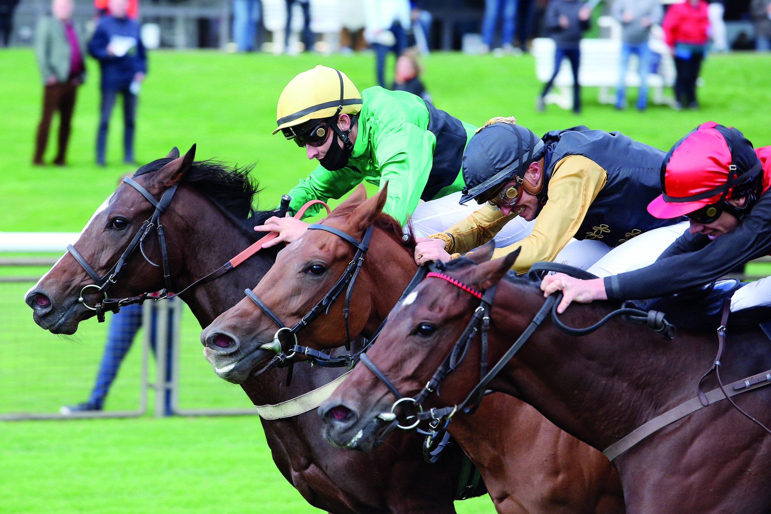
by Kate Allen and Geoffrey Lane
The majority of upper airway (‘wind’) disorders affect the regions of the pharynx and larynx. Most of these conditions are only present during exercise, when the upper airway is exposed to large changes in pressures associated with increased breathing rate and effort. This is the reason why performing endoscopy at rest may not give an accurate diagnosis. Endoscopy during strenuous exercise (overground endoscopy) has become key for veterinary surgeons to be able to give an accurate interpretation of upper airway function.
There are many different forms of upper airway disorders. They occur when part of the pharynx or larynx collapses into the airway, causing an obstruction to airflow. This obstruction causes turbulence to airflow, which in turn creates the abnormal noise. Observations of upper airway function during exercise enable veterinary surgeons to estimate the impact of the abnormalities with respect to race performance. Generally speaking, the more the structure collapses and the more the airway is narrowed, the greater the detrimental effect to performance. The mechanisms by which upper airway disorders affect performance are surprisingly complex, but in brief they influence the amount of air the horse can breathe in and also how hard the horse has to work to get that air into the lungs.
A full understanding of an individual horse’s upper airway function allows targeted treatments to be performed. Although the more common treatments have been included here for completeness, it is important for you to discuss individual horses with your own veterinary surgeon.
Understanding the anatomy is the first step to interpreting upper airway function during exercise. When looking at an endoscopic image, the left side of the horse is on the right side of the image as we look at it, and vice versa (figure 1).

Figure 1: Most disorders of the upper airway are named according to the structure that is collapsing. Therefore, understanding the anatomy of the airway will help to understand the individual conditions.
← Horse’s RIGHT side : Horse’s LEFT side →

Fig 2a

Fig 2b
With good upper airway function, we are looking for full abduction (which means opening) of the arytenoid cartilages while the vocal cords and aryepiglottic folds remain stable, and the epiglottis retains a curved shape; the soft palate and pharyngeal walls also remain stable. This gives a wide opening called the rima glottidis for air to enter the lungs (Figure 2 a, b, c).
Figure 2 a, b, Images showing good upper airway function.
Palatal instability and dorsal displacement of the soft palate
In the normal horse, the soft palate is positioned beneath the epiglottis. Palatal instability comprises billowing movement of the soft palate and often coincides with flattening of the shape of the epiglottis. The appearance of palatal instability can differ between horses (Figure 3 a, b, c). Palatal instability often causes an inspiratory noise.

Fig 3a

Fig 3b

Fig 3c
Figure 3 a, b, c: Images showing different types of palatal instability.
Dorsal displacement of the soft palate (DDSP) occurs when the free border of the soft palate becomes displaced and comes to lie above the epiglottis (Figure 4 a, b, c). In this displaced position, there is a substantial obstruction of the rima glottidis. Sudden onset ‘gurgling’ expiratory noises are characteristic of DDSP. Palatal instability almost invariably precedes DDSP, and it is thought these conditions may arise through weakness of the muscles within the palate itself.

Fig 4a

Fig 4b
Figure 4 a, b : Images showing dorsal displacement of the soft palate (DDSP). The epiglottis is no longer visible as the soft palate is now positioned on top of it.
Thus, in younger racehorses, palatal instability and DDSP will often improve with fitness and maturity. In the UK, the two most commonly performed surgical treatments are soft palate cautery and laryngeal tie-forward. The purpose of the soft palate cautery is to induce scar tissue to tighten the soft palate. The tie-forward has a different rationale. In some horses, the larynx slips backward just prior to DDSP, therefore the tie-forward holds the larynx in a more forward position, thereby inhibiting displacement.
Arytenoid cartilage collapse
This condition is also called recurrent laryngeal neuropathy, laryngeal hemiplegia or laryngeal paralysis because it is caused by nerve damage to the muscles of the larynx. During exercise, we observe collapse of the arytenoid cartilage almost always on the left side. In the context of sales, most trainers are familiar with laryngeal function grading applied during resting endoscopy. The purpose of this is to predict what is likely to happen to arytenoid function during exercise. During exercise, arytenoid function is typically graded as A, B or C where A is full abduction, B is partial collapse and C is complete collapse (Figure 5 a, b, c). The majority of horses with grade 1 or 2 laryngeal function at rest have grade A function during exercise (96% and 88% respectively). Arytenoid cartilage collapse causes a harsh inspiratory noise, often termed ‘roaring’.

Fig 5a

Fig 5b

Fig 5c
Figure 5 a, b, c: Images from 3 different racehorses, showing the variations in position of the left arytenoid. The first image shows a good position, followed by horses with increasing severity of collapse. In the last image, there is virtually no opening remaining for airflow.
Arytenoid cartilage collapse occurs when the nerve supply to the left side of the larynx is damaged. The most frequent surgery to improve complete collapse is a ‘tie-back’, which fixes the collapsing left side into a semi-open position. The potential limitation of this surgery is that if the arytenoid is fixed open, it cannot close to protect the rima glottidis during swallowing. Therefore, horses that have had a tie-back are susceptible to inhaling food into the lower airways leading to coughing. The tie-back is associated with a higher risk of complications than all other upper airway surgeries. More recently a nerve grafting surgery has been developed in which a normal local nerve is detached from a local muscle and then implanted into the laryngeal muscles. This avoids the potential complications of food inhalation but does take a few months to take effect. Both of these surgeries can be combined with ‘Hobday’ surgery.
Arytenoid Subluxation
This condition seems to be observed with increasing frequency. We see it most commonly in young flat racehorses; it is far less common in National Hunt horses, which probably reflects maturity of the laryngeal structures. One arytenoid subluxates or slips underneath the other arytenoid (Figure 6 a and b). The full name for this condition is ventromedial luxation of the apex of the corniculate process of the arytenoid cartilage (VLACPA). This condition appears to lead to instability of several other areas of the larynx, most commonly the vocal cords and aryepiglottic folds (Figure 7 a and b). There is limited scientific evidence for the best way to manage this disorder, and at present there is no effective surgical treatment. The instability within the larynx can be exacerbated the more the horse is exercised, therefore limiting the intensity of training to allow the larynx to mature may be recommended.

Fig 6a
Fig 6b
Figure 6 a and b: Images to show a closeup of the arytenoid cartilages. The image on the left is normal, and the two arytenoid cartilages meet in the middle. The image on the right shows that one side of the larynx has subluxated or slipped underneath the other side.

Fig 7a

Fig 7b
Figure 7 and b: Images to show arytenoid subluxation which has led to aryepiglottic fold collapse and vocal cord collapse.
Vocal cord collapse
Vocal cord collapse is often described as mild, moderate or severe, and typically causes a high-pitched inspiratory ‘whistle’ noise. Vocal cord collapse will almost always occur if arytenoid cartilage collapse occurs (Figure 8) but can also occur without arytenoid cartilage collapse (Figure 9). The traditional treatment for vocal cord collapse is the ‘Hobday’ procedure, which aims to remove the mucosal pocket to the side of the vocal cord along with the cord itself.

Figure 8: Image showing left arytenoid cartilage collapse with vocal cord collapse.

Figure 9: Image showing severe bilateral vocal cord collapse.
Aryepiglottic fold collapse
Aryepiglottic fold collapse is when the folds of tissue on the side of the larynx get sucked into the airway (Figure 10 a , b, c). This condition also causes a high-pitched inspiratory noise. It is typically graded as mild, moderate and severe. It most often occurs in conjunction with other conditions that alter the normal conformation of the arytenoid or epiglottis (i.e., palatal instability, arytenoid subluxation, arytenoid cartilage collapse). Treatment aims to remove a section of the folds.

Fig 10a

Fig 10b

Fig 10c
Figure 10 a, b, c: Images showing aryepiglottic fold collapse.
Pharyngeal wall collapse
Pharyngeal wall collapse is when the roof or sides of the pharynx collapse, which tends to obscure the larynx from clear view (Figure 11 a and b). It occurs more commonly in sport horses than racehorses due to head and neck position; the more flexed the head and neck position, the harder it is for the walls to remain stable. The time that we most often observe it in racehorses is at the start of the gallops if they are restrained, and often it will improve as the horse is able to extend its head and neck. This condition also causes a coarse inspiratory noise.

Fig 11a

Fig 11b
Figure 11 a and b: Images showing pharyngeal wall collapse.
Epiglottic entrapment
Although included here for completeness, epiglottic entrapment can usually be diagnosed during a resting endoscopic examination, particularly if the horse is triggered to swallow. The epiglottis becomes enveloped in the excess tissue that should lie underneath it (Figure 12 a and b). Sometimes the epiglottis remains entrapped, but sometimes it will entrap and release on its own which can make the diagnosis more difficult. The noise caused by epiglottic entrapment can vary, depending on the thickness of the entrapping tissue and whether DDSP occurs concurrently. Treatment involves releasing or resecting the excessive tissue.

Fig 12a

Fig 12b
Figure 12 a and b: Images showing epiglottic entrapment in two different horses. The image on the right shows an epiglottic entrapment that is more long standing, and the tissue has become swollen and ulcerated.
The disorders outlined above are described as if they are isolated single entities, but it is commonplace for horses to sustain complex collapse, which means collapse of multiple structures at the same time. Other less common disorders are epiglottic retroversion (when the epiglottis flips up to cover the rima glottidis), and cricotracheal membrane collapse (when there is collapse between the larynx and the trachea). On occasion obstructions to breathing can also occur in the nasal passages and the trachea (i.e., masses, ethmoid haematoma, sinusitis), but are far less common than those of the pharynx and larynx.
Looking forward it is unlikely that any new conditions remain to be discovered. Research now centres around better understanding of the causes of these disorders and how best to prevent and treat them. A particular area of investigation amongst several research groups is understanding how to train the upper airway muscles more appropriately to reduce the prevalence of these disorders and to investigate methods to strengthen the muscles. This would have the potential to reduce the number of horses needing surgical treatments.
The Biome of the Lung

By Dr. Emmanuelle van Erck-Westergren, DVM, PhD, ECEIM
Of bugs and horses
A couple of weeks ago, I was on an emergency call to a training stable. Half of the horses had started coughing overnight, some had fever, and, as you’d expect when bad karma decides to make a point, the two stars of the premises, due to face their greatest challenge to date the following week, were dull and depressed. A thick and yellow discharge was oozing from their noses. It was not long before the barn became the typical scene of a bad strangles nightmare. The bacteria involved in strangles outbreaks are Streptococcus equi equi, highly aggressive and contagious germs that spread fast and cause disruption in days of training and mayhem in tight racing schedules.
So what inevitably comes to mind when you hear the words “germs” or “bacteria”? Certainly no nice and friendly terms. As veterinarians, we have been taught that microorganisms are responsible for an endless list of gruesome diseases and conditions: abscesses, pneumonia, septicemia ... you name it. All of these need to be identified and eradicated. Thank heavens we still have an arsenal of antibiotics to get rid of the damn bugs. But recent research in human “microbiome” is making us think twice, especially as we aim to hit hard and large with antibiotics.
Never alone
Your healthy and thriving self, and likewise your horse, hosts millions and trillions of bacteria. The “microbiota” is that incredibly large collection of microorganisms that have elected you and your horse as their permanent home. The microbiota is constituted not only by an extremely diverse variety of resident bacteria, but also by viruses, fungi, and yeasts that multiply in every part of your external and internal anatomy. The discovery of this prosperous microbial community has triggered fascinating new research. It has unveiled the unsuspected links that exist between health, disease, and the microbiota. In simple words, these microorganisms are vital to your strength and healthiness.
The microbes that compose the microbiota outnumber our own cells by 10 to one to the extent that the genetic information (or “genome”) you carry is over 99% microbial! And that is what researchers call the “microbiome” or “biome”: the collection of genetic information carried by your microbiota. Fortunately, the very large majority of bacteria is either beneficial or harmless, with only a very tiny fringe represented by potentially pathogenic strains. These microorganisms have evolved with us over thousands of years and the stability of this symbiotic ecosystem has important implications on our health status.
A gut feeling for biome
Research on the biome started with the study of the digestive ecosystem of mice. Researchers from Washington University showed that when they transplanted feces of obese mice in the gut of lean mice, these became obese, and vice versa. In other words, the composition of the gut biome could be said to influence morbid weight gain. Similar studies recently conducted in humans in the Netherlands came to the same conclusions.
We do not yet have all the keys to understanding the underlying processes, but we definitely know that gut microbes influence, amongst many other things, our metabolism, which is to say our capacity to process energy. This opened up tremendous possibilities to improving fitness and treating diseases. The research on the biome has since grown at an exponential rate, covering much larger areas. It was further discovered that problems in the gut biome leading to the proliferation of the wrong microorganisms were responsible for a very wide range of disorders or even chronic conditions that were far from the gut, such as arthritis, depression, and asthma. The biome also seems to be critical in regulating our immune system to raise the alarm when enemies are identified and to modulate its response. The dramatic rise in autoimmune diseases could be a consequence of dietary changes that have disrupted our healthy microbiota.
TO READ MORE --
BUY THIS ISSUE IN PRINT OR DOWNLOAD -
August - October 2018, issue 49 (PRINT)
$5.95
August - October 2018, issue 49 (DOWNLOAD)
$3.99
Why not subscribe?
Don't miss out and subscribe to receive the next four issues!
Print & Online Subscription
$24.95
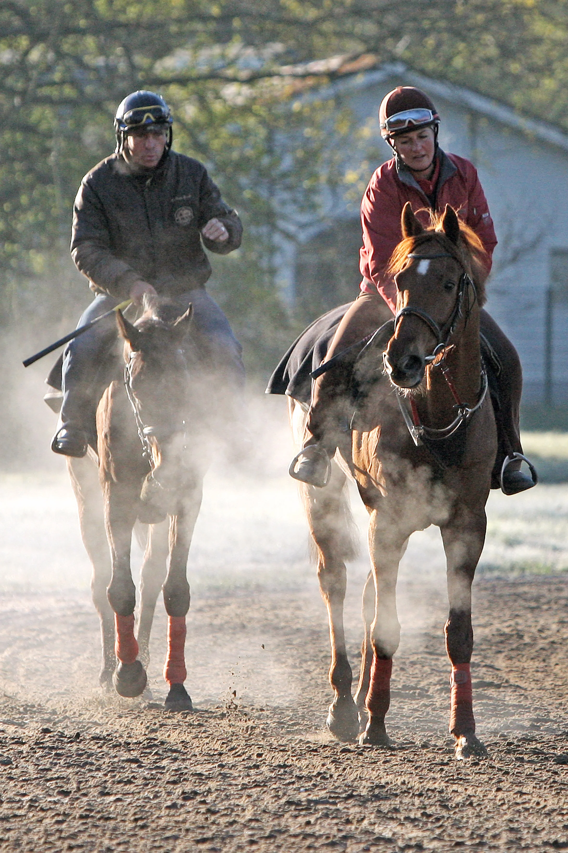
The Art of Breathing
Over the last two decades the Horserace Betting Levy Board (HBLB) in England has funded substantial research to understand how various body systems respond to training. For example, because of this HBLB investment we now know that the hearts of Thoroughbred racehorses get bigger as a response to athletic training and that big hearts are typically associated with better performers.

CLICK IMAGE TO READ FULL ARTICLE