The latest Strangles research available testing for ultimate sensitivity to avoid infection
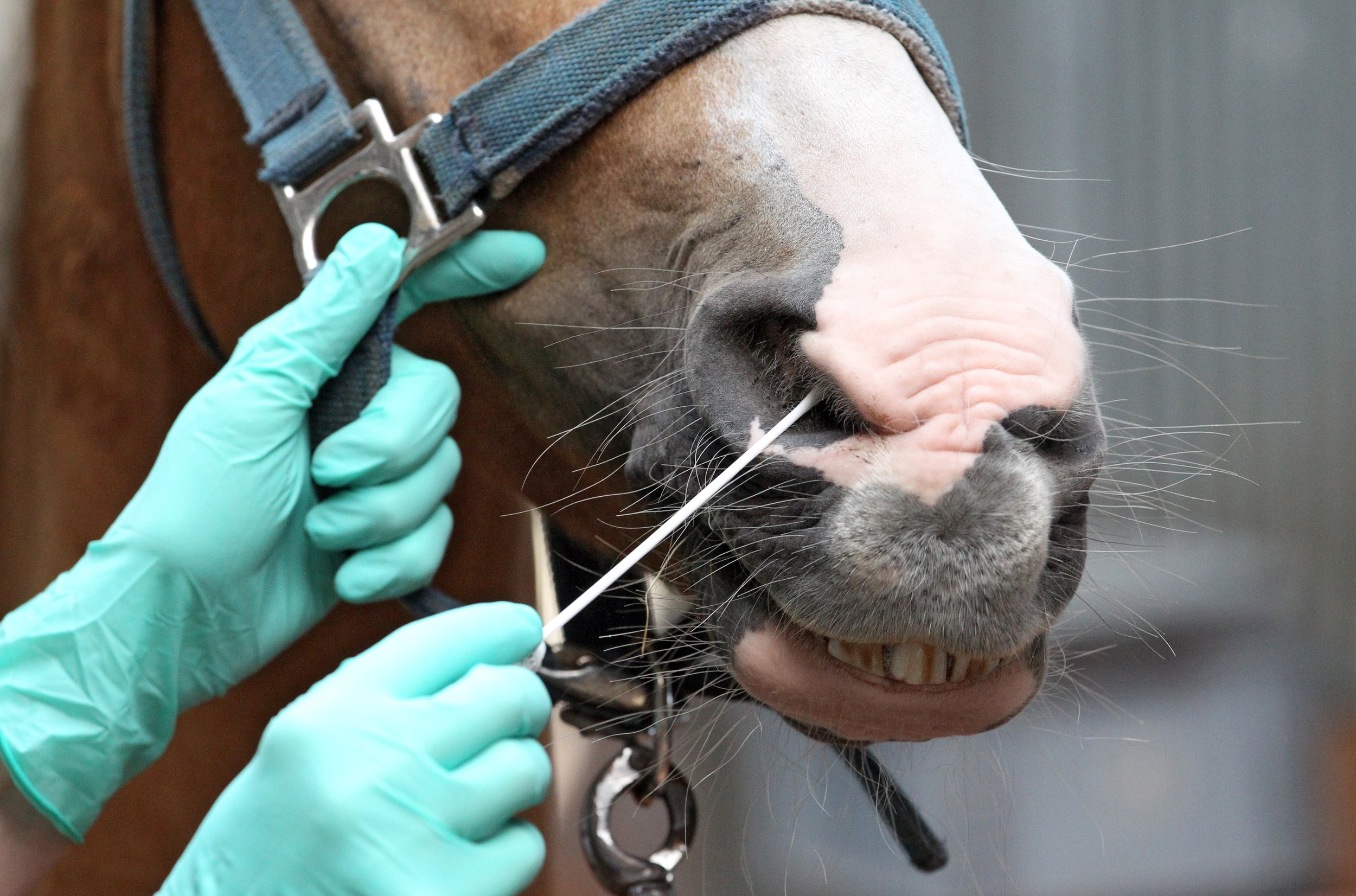
Strangles, the highly contagious upper respiratory disease caused by the bacterium, Streptococcus equi (S. equi) has been front and centre on social media lately with numerous disease alerts being posted. These alerts are triggered by positive test results for S. equi and reported by an official laboratory to the provincial or state veterinary office. Given the potential ramifications of a positive test, such as animal movement restrictions for several weeks and increased costs to horse and facility owners, a lot rides on the interpretation of these test results and the associated risk of disease spread to other horses, on and off the premises.
Testing for S. equi helps determine that a horse is free of S. equi or, in other words, not an S. equi carrier. It is usually done when the horse has recovered from clinical signs of Strangles to determine they are no longer infected and capable of transmitting S. equi, or upon request by equine facility managers, to screen a horse for carrier status prior to coming to their facility.

The two tests utilized for S. equi testing are the polymerase chain reaction (PCR) and bacterial culture. Testing utilizing bacterial culture detects living S. equi. Polymerase chain reaction (PCR) testing is much more sensitive than culture but detects DNA from both living and non-living bacteria. While the PCR sensitivity level can be useful as it can detect carrier horses that have a very low level of bacteria present in their guttural pouches, it can also detect transiently exposed/infected, asymptomatic horses, which rapidly clear the infection within a week. PCR can also flag horses that are less likely to be infectious at the time of sampling which can aid in risk management for that horse and the herd.

While these tests have their pros and cons, the relationship between S. equi PCR and bacterial culture has not been extensively studied. This is what Dr. Scott Weese of the Ontario Veterinary College and collaborators from OMAFRA and the University of Prince Edward Island set out to determine in a 2023 research study funded by Equine Guelph; ( tinyurl.com/guelph-strangles)
The relationship between quantitative real-time PCR cycle threshold and culture for detection of Streptococcus equi subspecies equi.
The 2023 study compared PCR and culture results from 158 equine respiratory tract samples submitted to an Ontario animal health laboratory for S. equi PCR testing. Of the samples that were PCR positive (CT < 40), only a minority (7.6%) were positive for S. equi on culture. That suggested that most PCR positive horses were likely a low risk for transmitting the bacterium at the time of sampling. A qPCR cycle threshold (CT ) of 34.2 was the breakpoint established, signifying that the likelihood of finding culturable S.equi above a CT of 34.2 was less likely and that the horse had a lower risk of being infectious at that point in time. These results were specific to this particular laboratory and cannot be applied to other laboratories which use their own testing procedures.
The line is not meant to be a green or red light but an indicator to aid in assessing the risk of disease transmission. Horses with PCR CT levels above 34.2, and who have developed a carrier status, can go on to produce lower CT levels (higher bacterial counts) over time and be a risk for S. equi shedding down the road. More research is needed to understand the S. equi shedding dynamics in carrier horses.

Combining culture and PCR testing is an option which comes at a higher cost to the horse owner but can be useful for an in-depth way to investigate bacterial loads and the risk of transmission at the time of sampling. While opting for ultimate sensitivity can help make sure no potentially infected horses are missed, it can start the domino effect of excessive control measures and costly interventions if not put into perspective related to the goals of testing, which may vary significantly between facilities (e.g. busy show barn, racetrack or closed herd).
Strangles has existed in horses since the 1800’s and isn’t going away anytime soon. Testing as part of a recovery plan from a Strangles outbreak is a no-brainer, but when it comes to using S. equi testing as part of a sickness prevention plan for your horse or facility, talk with your veterinarian and understand the impact a positive test result might have on your horse/herd and wallet BEFORE you start testing.
Electroarthrography to Predict Cartilage Quality
Article by Jackie Zions interviewing Dr. Adele Changoor and Dr. Judith Koenig

Researchers from the Ontario Veterinary College (OVC) and University of Toronto are developing a novel method to measure the quality of cartilage in horses using electroarthrography (EAG). EAG is a non-invasive technique that uses electrodes attached to the skin around a joint to detect electrical signals produced by the cartilage when it is loaded.
Dr. Adele Changoor, from the University of Toronto and Lunenfeld Tanenbaum Research Institute, and Ontario Veterinary College researcher Dr. Judith Koenig from the department of Clinical Studies, explain how EAG works and why it may become very useful for predicting cartilage quality and diagnosing osteoarthritis and other degenerative joints diseases in horses.
EAG is analogous to electrocardiography (ECG), which measures the electrical activity of the heart. Cartilage produces electrical signals during loading and these signals reflect its biomechanical properties, such as stiffness and permeability.
“By measuring EAG signals, we can get an idea of how healthy the cartilage is,” said Changoor.

Healthy cartilage ensures joints can move without pain and has an important role preventing wear and tear on bone.
Currently, there are no readily available tools to assess cartilage quality in horses with the exception of diagnostic arthroscopy – a minimal invasive surgery – under general anesthesia. X-rays and ultrasound are not sensitive enough to detect cartilage changes, and magnetic resonance imaging (MRI) is expensive, requires anesthesia and is often difficult to access. EAG offers a potential alternative that is fast, easy, and affordable.
“EAG is a promising tool for detecting cartilage damage early allowing intervention with treatments that can slow down or prevent further deterioration of the joint,” says Koenig “EAG could also help us monitor the effectiveness of treatments over time.”
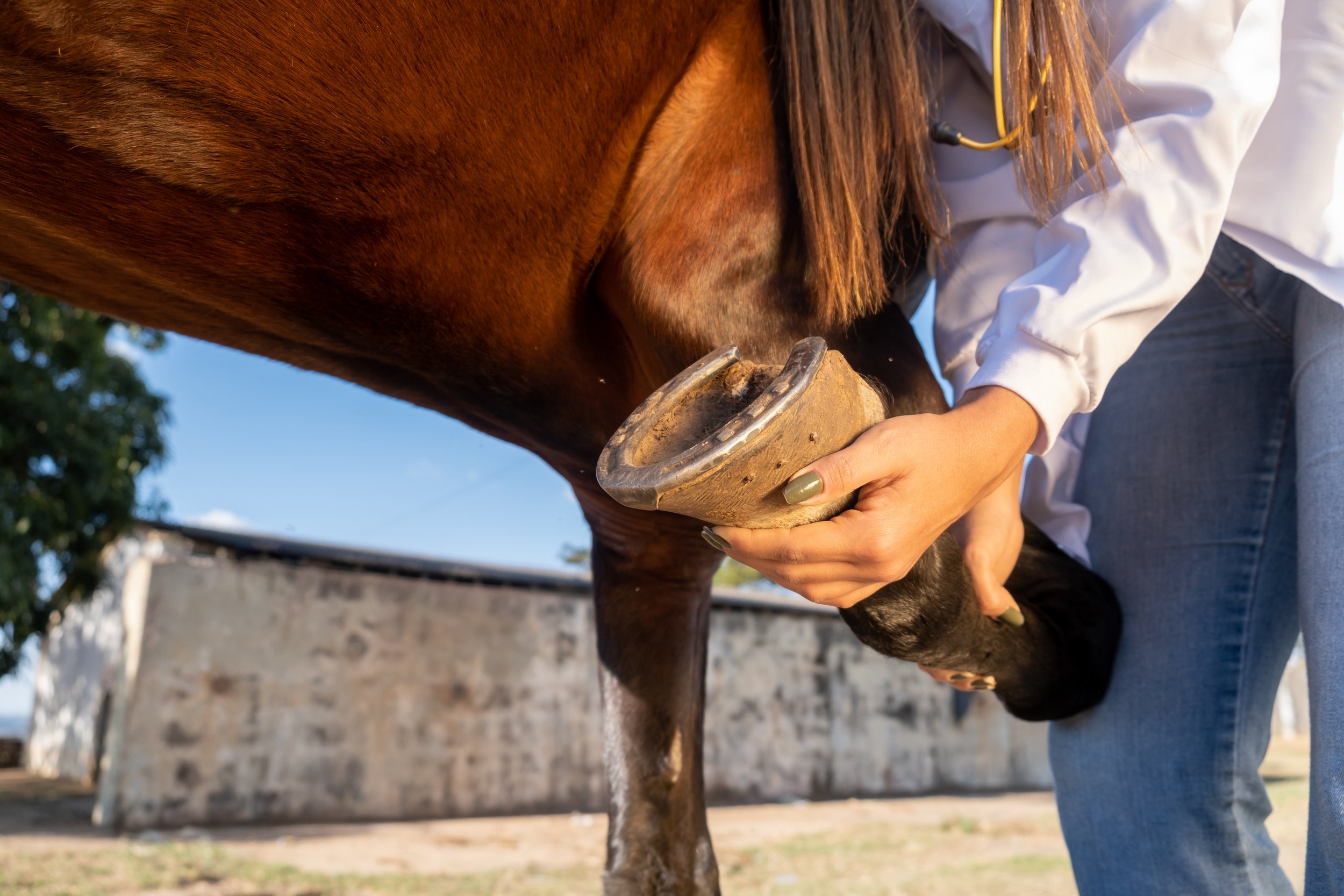
EAG measurements were collected at the same time as the center of pressure (COP), which measures the distribution of force under the horse’s hoof when it stands or walks.
“EAG is really tied directly to cartilage biomechanical properties,” says Changoor. “We also needed to know about the joint biomechanics in order to interpret EAG properly.” A custom, portable, force mat was developed by Dr. Changoor’s graduate students that included an array of force sensors to place under the horse’s hoof when measuring EAG.
“Then we can measure how much compressive force or ground reaction force is being exerted on that joint” said Changoor. “COP, is where the ground reaction force is acting. The ground reaction force gives us the total load on the joint. COP lets us figure out where on the hoof or where on the joint surface force is being concentrated.”

COP provides information about the joint biomechanics and the horse’s balance and stability. EAG and COP testing were combined to get a comprehensive picture of the joint health and function in horses with osteoarthritis. Results were compared with MRI imaging and it was found that EAG and COP testing correlated well with MRI and could detect differences in cartilage quality between healthy and osteoarthritic joints.

In the 2023 study involving horses with osteoarthritis in the fetlock joint; the horses were treated with MSCs to decrease inflammation and stimulate tissue healing. The researchers measured EAG, COP, and MRI before and after the treatment to evaluate its impact on cartilage quality.
“We observed that MSCs improved cartilage quality in some horses and EAG and COP testing were able to capture these changes and show the responses to treatment. This suggests that EAG and COP testing could be useful for selecting treatment options for the horse,” says Dr. Koenig. “One of the biggest advantages of EAG is that it seems to correspond with our arthroscopic findings. It can perhaps evaluate the quality of the cartilage or cartilage defects, which we are at the moment only able to evaluate with arthroscopy.”
The researchers plan to conduct further studies in order to validate and refine EAG and COP testing for predicting cartilage quality in equines. They hope that these techniques will become widely available and accessible for veterinarians and horse owners in the future.
“This is an exciting and innovative research project that has the potential to improve the diagnosis and early management of osteoarthritis in horses,” says Dr. Koenig “Osteoarthritis is a major health and welfare issue for horses and their owners, and we need better tools to detect it early and treat it. EAG and COP testing could provide a simple, affordable, and accurate way to assess cartilage quality and joint function in horses.”
Many thanks go to the graduate students who worked tirelessly on the EAG study: Peter Suderman, PhD Candidate in the Department of Materials Science & Engineering at U of T, Jaylon Pascual, undergraduate co-op student finishing her fourth year in the Biomedical Engineering program at U of G, Dr Rodrigo Munevar Luque, Equine Sports Medicine Resident at OVC and PhD Candidate Biomedical Sciences at U of G, Undergraduate Research Assistants in Clinical Studies Ashley Nixon, DVM 25 (OVC) , Pjotr Roest DVM 26 (OVC), and in Biomedical Sciences Axel Koenig Parris HBA 25 (Ivey School of Business, Western University) and Rebecca Mullin BSc OVC 25.
The study was funded by the Equine Guelph Research Fund and the Natural Sciences and Engineering Research Council of Canada (NSERC).
Gut issue biomarkers and their use in signalling dysbiosis
Article by Jackie Zions

Gastrointestinal issues (GI) are the number one cause of morbidity in horses other than old age. An unhealthy digestive system can cause poor performance, pain, discomfort, diarrhea, and a whole host of issues that can sideline your horse. It’s no wonder researchers are paying close attention to the ‘second brain’ and it’s billions of inhabitants. Ontario Veterinary College (OVC) researcher, Dr. Luis Arroyo has been studying the equine gastrointestinal systems for many years with several research projects receiving funding from Equine Guelph. Arroyo discusses what we know about equine gut health, causes of GI disorders and the extensive continuing research to understand what unstable and stable gut populations look like.
Starting with some basic anatomy Arroyo says, “The gastrointestinal tract of a horse is extremely large, and there are many things that can cause disturbances to the normal functioning or health of the gut.” A healthy gut microbiome is essential for the horse’s entire body to function optimally.
Signs of GI issues

Common signs of disorders could include abdominal pain, bloating, changes in fecal consistency (including diarrhea or constipation), excessive drooling, decrease in water consumption, lack of or poor appetite, weight loss and low body condition score.
“Some cases are more obvious to owners,” says Arroyo, “like poor performance, or acute or chronic diarrhea.”
Changes of behaviour such as becoming cranky or moody can be tell-tale signs there is unrest in the GI system. Biting at the flanks can signal abdominal pain as well as reactivity to being saddled. When the horse stops wanting to perform and athletic abilities suddenly decline, if there is no obvious lameness, GI issues are high among the considerations.
“Horses are herbivores, designed to consume a diet of forage, and to break down complex sugars within that forage.” says Arroyo. “The gut microbiota does this job and is very important for healthy digestion.” Recent research is connecting the changes in diversity of microbial communities to conditions like colic, colitis, and gastric ulcers.
Causes of GI Issues
Colic is the number one clinical condition occurring in horses. It is well-known that sudden dietary changes can be a major contributor as well as diets that are high in grain. This can create changes in the volatile fatty acids produced in the GI system, which in turn can lead to the development of gas colic. Arroyo provides the example of switching from dry hay fed in the winter, too rich, lush, spring grass as a big cause of rapid fermentation that can cause colic.

Any abrupt change, even if it’s a good quality feed to a different good quality feed, can be a source of colic. Then there is the more obvious consumption of moldy, poor, quality hay. So not only the quality but the transition/adaptation period needs to be considered when making feed changes and this goes for both changes to forage or concentrates.
A table of feed transition periods on the Equine Guelph website states an adaptation period of at least 10 – 14 days is recommended. Transition periods under seven days can increase colic risk over 22 times!
“Decrease in water consumption can be an issue, especially in countries with seasons,” says Arroyo. When water gets really cold, horses often drink less, and if it freezes, they don’t drink at all, which can lead to impaction colic. Parasite burden can also cause colic. If your horse lives in a sandy environment, like California, ingesting sand can cause impaction colic.
Non-steroidal anti-inflammatory drugs (NSAIDS) can cause colic or ulcers. NSAIDS can interfere with blood supply to the GI tract causing ulceration, for example in the mucosa of the stomach. Prolonged use can cause quite severe ulceration.
NSAIDS are not the only drugs that can contribute to GI issues. “Antibiotics - as the name says - kill many kinds of bacteria,” says Arroyo. “They are designed for that! Invariably they deplete some bacterial populations including in the intestine, and that is a problem because that may allow some other bacteria, potentially pathogenic or harmful, to overgrow, and that can cause dysbiosis.”
In a recent study, by fellow OVC researcher, Dr. Gomez and co-workers, it was determined that damage to the intestinal microbiota could occur after only 5 days of administering antibiotics to horses. Damage to the intestinal microbiota resembled dysbiosis that can potentially result in intestinal inflammation and colitis predisposing the horse to diarrhea. Judicious use of antibiotics and antimicrobials are advised.
There are infectious and non-infectious causes of colitis. Infectious examples include salmonella and then there is Neorickettsia risticii, which if ingested from contaminated sources, can cause Salmonellosis or Potomac horse fever, respectively.
“Any stress factors such as transportation, fasting or intense exercise like racing, can be a factor for developing stomach ulcers,” says Arroyo.
Current Diagnostics
Putting together a picture of the horse’s health status includes gathering clinical history from the horse owner and performing a physical examination for motility and hydration status. A biochemistry profile and complete set count can be gathered from blood testing.
Gastric ultrasound allows veterinarians to view the wall of the intestine, noting if it has thickened or distended, which could occur in cases when there is colic. They can assess appearance and find out if the intestine is displaced or if there is a twist. Gastroscopy is commonly used to find ulcers in the stomach and can reach as far as the first part of the duodenum.
GI Research

“DNA sequencing has been a breakthrough in science in terms of understanding the communities of different microorganisms living in many different niches from the skin to the lungs to the upper airways to the intestine,” says Arroyo.
It has allowed in-depth study of the population of microorganisms, providing a big picture of the different inhabitants in various areas of the GI tract, such as the lumen of the small intestine and the small and large colon. “The microorganisms vary, and they have different functions in each compartment,” says Arroyo.
DNA sequencing has allowed researchers to study microbial populations and gather information on what happens to bacterial communities when impacted by diseases like colitis. “We can see who is down, and who is up,” explains Arroyo, “and determine what populations have been depleted.” It has led to a better knowledge of which of the billions of factors are harmful to the system and which can compromise the health of the horse.
Robo-gut is one example of a fantastic system where bacterial communities are being replicated in the lab to mimic what would be found in a natural environment.
Researchers at the University of Guelph have measured metabolic profiles of the bacterial population after the addition of supplements like probiotics and prebiotics. They found they can dramatically change the metabolites that are being produced, according to what is being added to the system.
Exciting new research that could impact the future of diagnostics includes screening for biomarkers as indicators of intestinal health among equine microbiota. Dr. Arroyo is currently working with research partner, Dr. Marcio Costa, from the University of Montreal, looking for biomarkers that indicate changes in the inhabitants of the equine gut that take place during the early onset of illness.
“A biomarker is a biological molecule that you can find in different places,” explains Arroyo. “For example, you might find them in tissue, blood, urine, or different body fluids. They can signal normal or abnormal processes or could reveal a marker of a disease. For example, a biomarker can be used to see how well the body might respond to a treatment or to a disease condition.”
“The objective of a dysbiosis index is quantifying ‘X’ number of certain bacteria that are important to us,” says Arroyo. In this case, the dysbiosis derives from sequencing of the bacterial population in fecal samples.

Changes in the intestinal microbiota (dysbiosis) are present before and during the outset of diseases and after treatment with antibiotics. Arroyo cites the example of decreased Lachnospiraceae commonly observed when there is intestinal inflammation.
Bacterial biomarkers are currently being used in other species to accurately predict intestinal dysbiosis, for example in cats and dogs. One canine study quantified the number of seven different taxa of importance of the total bacterial populations. This information is entered into a mathematical algorithm that comes up with results explaining which bacteria have increased or decreased. Based on those numbers, one can use a more specific taxa to identify dysbiosis. In a feline study, it was discovered that six bacterial taxa could be accurately used to predict diarrhea in 83% of cases.
It is hoped the same results could be accomplished for horses. Developing PCR testing to screen for biomarkers could be a game changer that could potentially provide speedy, economical early diagnostics and early treatment.
So far, the most remarkable finding in the preliminary data reveals that in horses with colitis, the whole bacterial population is very depleted.
“At this stage we are in the process of increasing our numbers to find significant differences in which bacterial taxa are more important,” says Arroyo. “Soon we hope to share which bacteria taxa are more promising for predicting dysbiosis in horses with gastrointestinal disease.”
The researchers are delving into a huge biobank of samples to identify potential markers of intestinal dysbiosis in horses, utilizing PCR testing as a faster and more economical alternative to the complex DNA sequencing technologies that have been used to characterize changes in microbiota thus far. The goal is to develop simple and reliable testing that veterinarians can take right to the barn that will result in early treatment and allow closer monitoring of horses at the first onset of GI disease.
Top Tips to Protect Digestive Health

Horses are hind gut fermenters who rely on adequate amounts of fiber in the diet to maintain healthy gut function.
Make dietary changes slowly as abrupt changes disrupt the microbiota.
Avoid large grain meals as huge portions of highly fermentable diets can be quite harmful to the microbiota and can also be a source of risk for developing gastric ulcers. Opt to spread out concentrates into several smaller rations.
Prevent long periods of fasting which can also lead to ulcers. Horses are continuous-grazers, and they need to have small amounts of feed working through their digestive system to keep it functioning optimally.
Have a parasite prevention program.
Provide fresh water 24/7 to maintain good hydration and keep contents moving smoothly through the GI tract.
Keep up to date on dental appointments.
Motion is lotion – turn out and exercise are extremely important to gut function.
In closing, Arroyo states, “These top tips will help keep the horse happy and the gastrointestinal tract functioning properly.”
