Electroarthrography to Predict Cartilage Quality
Article by Jackie Zions interviewing Dr. Adele Changoor and Dr. Judith Koenig

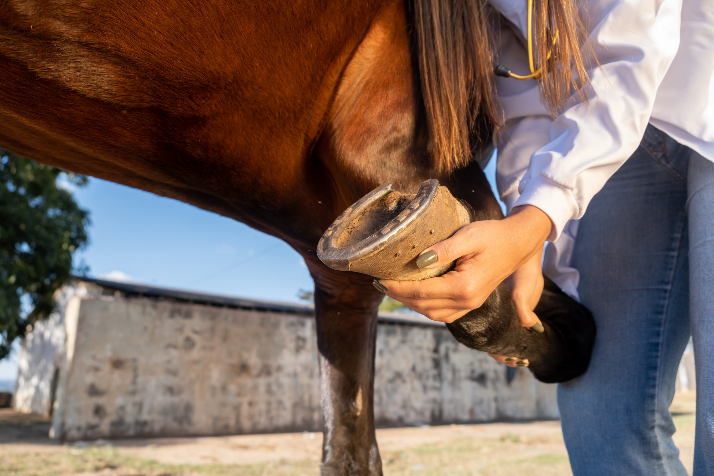
Researchers from the Ontario Veterinary College (OVC) and University of Toronto are developing a novel method to measure the quality of cartilage in horses using electroarthrography (EAG). EAG is a non-invasive technique that uses electrodes attached to the skin around a joint to detect electrical signals produced by the cartilage when it is loaded.
Dr. Adele Changoor, from the University of Toronto and Lunenfeld Tanenbaum Research Institute, and Ontario Veterinary College researcher Dr. Judith Koenig from the department of Clinical Studies, explain how EAG works and why it may become very useful for predicting cartilage quality and diagnosing osteoarthritis and other degenerative joints diseases in horses.
EAG is analogous to electrocardiography (ECG), which measures the electrical activity of the heart. Cartilage produces electrical signals during loading and these signals reflect its biomechanical properties, such as stiffness and permeability.
“By measuring EAG signals, we can get an idea of how healthy the cartilage is,” said Changoor.

Healthy cartilage ensures joints can move without pain and has an important role preventing wear and tear on bone.
Currently, there are no readily available tools to assess cartilage quality in horses with the exception of diagnostic arthroscopy – a minimal invasive surgery – under general anesthesia. X-rays and ultrasound are not sensitive enough to detect cartilage changes, and magnetic resonance imaging (MRI) is expensive, requires anesthesia and is often difficult to access. EAG offers a potential alternative that is fast, easy, and affordable.
“EAG is a promising tool for detecting cartilage damage early allowing intervention with treatments that can slow down or prevent further deterioration of the joint,” says Koenig “EAG could also help us monitor the effectiveness of treatments over time.”
EAG measurements were collected at the same time as the center of pressure (COP), which measures the distribution of force under the horse’s hoof when it stands or walks.
“EAG is really tied directly to cartilage biomechanical properties,” says Changoor. “We also needed to know about the joint biomechanics in order to interpret EAG properly.” A custom, portable, force mat was developed by Dr. Changoor’s graduate students that included an array of force sensors to place under the horse’s hoof when measuring EAG.
“Then we can measure how much compressive force or ground reaction force is being exerted on that joint” said Changoor. “COP, is where the ground reaction force is acting. The ground reaction force gives us the total load on the joint. COP lets us figure out where on the hoof or where on the joint surface force is being concentrated.”

COP provides information about the joint biomechanics and the horse’s balance and stability. EAG and COP testing were combined to get a comprehensive picture of the joint health and function in horses with osteoarthritis. Results were compared with MRI imaging and it was found that EAG and COP testing correlated well with MRI and could detect differences in cartilage quality between healthy and osteoarthritic joints.

In the 2023 study involving horses with osteoarthritis in the fetlock joint; the horses were treated with MSCs to decrease inflammation and stimulate tissue healing. The researchers measured EAG, COP, and MRI before and after the treatment to evaluate its impact on cartilage quality.
“We observed that MSCs improved cartilage quality in some horses and EAG and COP testing were able to capture these changes and show the responses to treatment. This suggests that EAG and COP testing could be useful for selecting treatment options for the horse,” says Dr. Koenig. “One of the biggest advantages of EAG is that it seems to correspond with our arthroscopic findings. It can perhaps evaluate the quality of the cartilage or cartilage defects, which we are at the moment only able to evaluate with arthroscopy.”
The researchers plan to conduct further studies in order to validate and refine EAG and COP testing for predicting cartilage quality in equines. They hope that these techniques will become widely available and accessible for veterinarians and horse owners in the future.
“This is an exciting and innovative research project that has the potential to improve the diagnosis and early management of osteoarthritis in horses,” says Dr. Koenig “Osteoarthritis is a major health and welfare issue for horses and their owners, and we need better tools to detect it early and treat it. EAG and COP testing could provide a simple, affordable, and accurate way to assess cartilage quality and joint function in horses.”
Many thanks go to the graduate students who worked tirelessly on the EAG study: Peter Suderman, PhD Candidate in the Department of Materials Science & Engineering at U of T, Jaylon Pascual, undergraduate co-op student finishing her fourth year in the Biomedical Engineering program at U of G, Dr Rodrigo Munevar Luque, Equine Sports Medicine Resident at OVC and PhD Candidate Biomedical Sciences at U of G, Undergraduate Research Assistants in Clinical Studies Ashley Nixon, DVM 25 (OVC) , Pjotr Roest DVM 26 (OVC), and in Biomedical Sciences Axel Koenig Parris HBA 25 (Ivey School of Business, Western University) and Rebecca Mullin BSc OVC 25.
The study was funded by the Equine Guelph Research Fund and the Natural Sciences and Engineering Research Council of Canada (NSERC).
Lower limb anatomy and how it can be conditioned for racing
Words - Adam Jackson MRCVS

Better understanding the appropriate levels of exercise and training while the horse’s body grows and develops has been a topic of research for many years. Although it has been shown that young, growing horses are well-suited to adapt to conditioning, it is vital that continued research is performed in order to develop thoughtful and strategic training methods to promote healthy, fit and sound horses with long careers and lives.
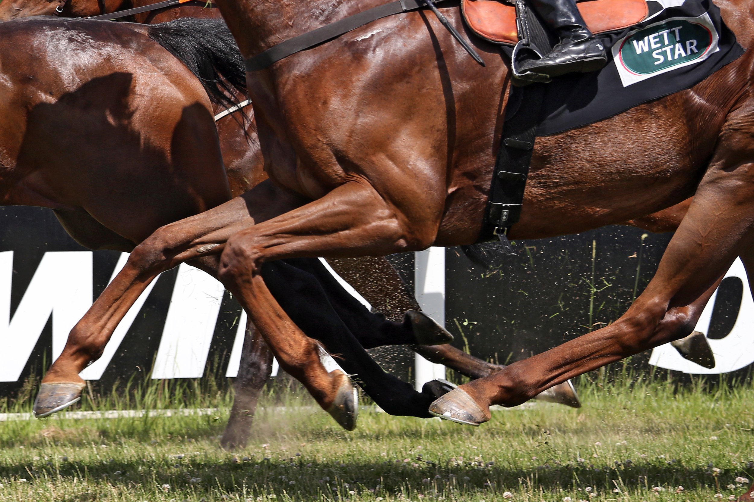
Horses’ limbs consist of dozens of muscles, bones, tendons, ligaments, and joints that allow the horse to move as well as support its body weight. The limbs function to provide thrust and movement while absorbing impact and bearing weight. Most of the horse’s weight is supported by the fore limbs, while the propulsion of the horse is provided by the hind limbs. In addition, the horse has two apparatuses referred to as the stay apparatus and suspensory apparatus. The stay apparatus allows major joints in the limbs to lock so that the horse may rest and relax while standing. The suspensory apparatus is designed to absorb shock, carry the horse’s weight, and prevent the overextension of joints. Finally, the hooves are important structures that maintain support and traction as well as provide additional shock absorption.
Since the cardiovascular system provides blood supply throughout the body, by responding to various stimuli, it can control the velocity and amount of blood carried through the vessels, thus, delivering oxygen, nutrients, hormones, and other important substances to cells and organs in the body. It plays a very important role in meeting the body’s demands during exercise, stress, and activity.
Exercise is used to increase the body’s ability to withstand repeated bouts of similar exercise with less impact. With a strong and healthy cardiovascular system, there is an improved ability of the musculoskeletal system receiving oxygen, thus, allowing muscles to better their capacity to use oxygen and energy. However, the adaptation period for each of these physiological systems do differ as the cardiovascular system adapts faster compared to the musculoskeletal system. This is often an overlooked consideration when developing training programmes for horses.
It is important to understand the various functions, structures, and adaptive processes of the horse’s musculoskeletal system such as bone, articular cartilage, tendons, and ligaments in order to develop appropriate training regimens.

Bone has many important roles that involve locomotion, the storage of minerals (especially calcium and phosphate), soft tissue and vital organ protection, and the support and containment of bone marrow. Bone is a specialized connective tissue, and together with cartilage forms the strong and rigid endoskeleton. The bone is continuously altering through two processes called bone modeling and bone remodeling, involving four cells referred to as osteoclasts, osteoblasts, osteocytes and bone lining cells.
Osteoblasts secrete bone matrix in the form of non-mineralized osteoid, which is then mineralized over a few weeks to form a bone matrix. Osteoclasts are involved in resorption of bone as this process occurs faster than the formation of bone. When the bone surfaces are not in the development or resorption phase, the bone surface is completely lined by a layer of flattened and elongated cells termed bone-lining cells. Osteocytes are derived from osteoblasts and are highly specialized to maintain the bone matrix. They are designed to survive hypoxic conditions and maintain biomineralization of the bone matrix. Osteocytes also control osteoblastic and osteoclastic activities allowing bone remodeling.
The function of bone modeling is to alter and maintain shape during growth. As the horse grows and develops, bone modeling occurs with the acquisition and removal of bone. While the young horse grows and develops, bone modeling allows the bone to endure strains from everyday work and exercise. The adult skeleton undergoes a minimal amount of bone modeling. Due to the presence of the high frequency of bone modeling in young horses, their skeletal strength is highly influenced by strains to their bones during exercise and daily use. With this knowledge, it has been concluded and confirmed that short-term dynamic exercise of an adolescent can lead to beneficial changes to its bone morphology.
Bone remodeling is a different process, in which old and damaged bone is renewed, which enables the bone to respond and adapt to changing functional situations. Bone remodeling is usually a coordinated relationship between bone resorption and bone formation. This process occurs throughout the horse’s life with the renewal of primary, damaged or old bone. Osteoclasts absorb old and damaged bone, and the osteoblasts form new bone and lay down new bone matrix until the earlier absorbed bone is replaced. In those animals with musculoskeletal disease or damage, there is an imbalance of osteoblast and osteoclast activity. With the knowledge that osteoblast activity to make new bone takes months whilst osteoclast activity of removing old and damaged bone only takes a few days to two weeks, bone that is being repaired is at a high risk of further injury as bone removed has not been completely replaced. Multiple studies have shown that exercise while growing can provide lifelong benefits; however, it must be done with care and knowledge. In addition, many studies have shown that exercise of a dynamic nature in moderate distances, such as that achieved in the pasture or prescribed short-distance high-speed work is beneficial to musculoskeletal development and may prevent injuries when entering race training. It has also been observed that long slow work does not increase bone strength. Below is a summary of the young horse response of the various types of exercise.

Articular cartilage is a highly specialized connective tissue found in joints with the role of providing a smooth, lubricated surface of articulation and to help transmit loads with a low amount of friction. The articular cartilage is a hyaline cartilage (flexible and strong tissue providing a smooth, slippery surface) with a dense “ExtraCellular Matrix” (ECM) consisting of specialized cells called chondrocytes, collagen and proteoglycans. These components help to retain water in the ECM that is required for the joints mechanical properties. As age increases, hydration of the matrix does decrease, resulting in stiffness. Chondrocytes are residential cells in articular cartilage that play a role in the development, maintenance, and repair of the ECM. They do respond to a variety of stimuli, including mechanical loads, growth factors, hydrostatic pressures, piezoelectric forces (formation of electric charge with force). Because of the lack of blood vessels, lymphatics, and nerves as well as being a harsh biomechanical environment, there is a limited capacity to heal and repair. In addition, chondrocytes have limited potential for replication, thus, have limited healing capacity; and chondrocytes survival depends on an optimal chemical and mechanical environment.
Maintaining joint health is vital, which requires the preservation of healthy cartilage tissue. Inactivity of joints is detrimental to articular cartilage; thus, regular movement of joints and dynamic loads is needed to provide a normal articular cartilage structure and function. Biochemical responses of the cartilage to exercise are not nearly as well known compared to bone. While the confinement of young horses stunts joint development, excessive straining of cartilage can also reduce joint development. It has been observed that pasture access was optimal for the development of joints and the confinement or excessive sprint exercise (12–32 sprints of 40 meters for 6 days a week for 5 months) causes detrimental effects on the joint and may be deemed as unnatural exercise. It is also thought that exercise is needed well before two years of age to allow cartilage thickening as well as the avoidance of confinement. It can be concluded that further studies are required with respect to level of exercise and type of exercise in order to achieve healthy cartilage tissue as there is clearly a fine line between frequency and intensity of exercise.

Tendons and ligaments are distinct but closely related tissues that have unique and important roles in musculoskeletal function and musculoskeletal disease. Tendons and ligaments are dense, fibrous connective tissues that connect muscle to bone or bone to bone, respectively. These tissues transmit mechanical forces to stabilize the skeleton and allow body movement. Tendons and ligaments consist mainly of collagen type I as well as small amounts of collagen III, IV, V, and VI. There are also various proteoglycans in tendons and ligaments that both organize and lubricate collagen fiber bundles. The elasticity of tendons and ligaments is due to the large amount of type I collagen. During locomotion, the tendon decreases energy cost to the horse by acting as a spring to store and release energy while stretching and recoiling in the stance and swing phases of each stride. Tendons and ligaments have blood vessels and nerves that allow the homeostasis and response to injury.
Tenocytes are tightly regulated by a series of growth factors and transcription factors that allow the synthesis, maintenance, and the degradation of the tendon extracellular matrix. Tendons are elastic, but tearing may occur if there is excessive loading on the tendon and the repair of collagen is a slow process. In addition, tendons have crimp morphology where the tendons buckle in a state of relaxation and act as shock absorbers. Unbuckling of the tendon occurs during loading. This crimp morphology may be disturbed if an injury occurs and also is reduced in older horses.
Due to the variation of activity of tenocytes in foals and young horses, it has been observed that both a lack of exercise and excess of exercise can impair tendon make-up and subsequent functionality. With the current data and research that has been gathered, it can be concluded that if horses take advantage of spontaneous exercise when in the paddocks (which they often do), the developing tendons may benefit and be at a lower risk of injury when racing training starts.
Conclusion
It is clear that further research is needed in order to ascertain the optimal amount and type of exercise that is needed in order to provide a strong musculoskeletal system and functional performance. However, it has been shown that prescribed exercise during the growth of the horse can increase the longevity of the horse’s health and performance. It has been observed that confinement and the lack of loading can result in weaker tissues and the loss of function of none, tendons, ligaments and articular cartilage. However, it must also be recognized that medical attempts to alleviate pain so that a horse can continue to train through an injury can greatly increase tissue damage which is detrimental to the horse’s health and career. It is far more beneficial to provide an adequate amount of time for the injury to heal, thus, putting the horse’s health and wellbeing as a top priority.
Nutritional perspective
Bone development in yearlings from the sales ring to racing — nutritional perspective
Words - Des Cronin B.Ag.Sc, M.B.A

Maintaining the equine skeleton is vital to ensure optimal development of the young growing horse, minimize risk of injury in the performance horse, and promote longevity and soundness.
The skeletal development and health of a young horse begins in utero and ensuring the broodmare receives the correct intake of key nutrients will be critical to the growth of the unborn foal. Producing high-quality milk places a significant drain on the mineral reserves of the mare. Maintaining mineral intakes during peak lactation is vital to ensure the foal receives the best nutrition to support the rapid skeletal development in the early weeks and months of growth. During this time, bone formation, body size, and muscle mass greatly increase. Risk of defective bone and related tissue formation increases with one of more of the following:
Poor diet with the incorrect balance of energy and nutrients in the daily ration
Inadequate amounts of calcium (Ca) and phosphorus (P)
A reversed Ca:P ratio
Low zinc (Zn) or copper (Cu) in the diet
Low Vitamin D
Feeding a young horse for a maximum growth rate is undesirable because bone hardening lags greatly behind bone lengthening. At 12 months old, the young horse could reach about 90 to 95 per cent of its mature height but only about 75 per cent of its mature bone mineral content.
Ideally, young horses should gain weight at a rate that their developing bones can easily support. Growing bones and connective tissues don’t have the strength to support rapid weight gain from overfeeding, especially energy. Rapid weight gain can also make other skeletal anomalies worse. In these cases the risk of developmental orthopedic disorders (DOD) and unsoundness increases.
DOD and unsoundness can also occur during uneven growth. For example, switching an underfed, slow-growing horse to a good diet that allows quick growth (compensatory growth), increases the risk of DOD. Foals between the ages of 3 and 9 months of age are at greatest risk of DOD.
Fresh forages, for example grazed grass, usually provide enough major minerals such as calcium (Ca) and phosphorus (P) for the growing horse. However, there can be significant variation in calcium and phosphorus levels in all forages but particularly preserved forages (hay and haylage). Forage analysis should always be undertaken to determine mineral composition.
For young fast-growing horses, the diet must supply the quantities of calcium and phosphorus needed for normal bone formation. In terms of Ca:P ratio, the ratio must be positive in favor of calcium. Horses are much more tolerant of high-dietary calcium than other species. For practical purposes, a good guideline would be to keep the ratio Ca:P between 1.5 to 1 and 2.5 to 1. Grains (e.g., oats) contain 10 per cent of the calcium level found in typical forages. Grains are poor sources of calcium, both in terms of the amount of calcium supplied and their effect on Ca:P ratio in the diet. Where grains are fed, supplementation will be necessary to balance the diet.
While some forages may contain adequate calcium and phosphorus, they will typically supply less than 20 per cent of the daily requirements for trace elements. Supplementation of trace elements will generally be necessary to support normal bone development.
Where concentrates are fed (especially low levels), supplementation may still be necessary to balance the overall mineral and trace element intake. Nutritional advice should be sought to ensure the horse's diet is correctly balanced.
To meet the carefully balanced requirements of key minerals, it is advisable to supplement the daily rations of growing horses and young horses entering training with an appropriate nutritional product.
Make sure that the supplement used contains the correct ratio of calcium and phosphorus, as well as other key nutrients such as vitamin D and chelated trace elements (copper, manganese, and zinc) to support normal bone development.
Supplementing branch chain amino acids in the diet ensures that growth is maintained. Lysine plays a key role when protein concentrations in the body are low. Vitamin A supports collagen formation, which is a key component of the supportive structures of joints (tendons and ligaments). Vitamin D3 is added to enhance calcium absorption.
Although growth rates slow after the age of two, they are still juvenile in their skeletal development with some growth plates, such as the shoulder and stifles, yet to completely close. Although they may look like fully grown adults, it is still important to meet nutritional requirements especially if starting training and work. With the addition of exercise and training, a young horse's nutritional needs change. The added forces from groundwork on the long bones and increased requirements of other nutrients like electrolytes need to be considered.
Finally, horses all grow and develop at different rates because of factors such as genetics. Some youngsters will need more support for longer periods of time than others, so it is important to manage accordingly.